Cell communication (biology)
Cell communication is the ability of cells to communicate with adjacent cells within an organism. The term is mostly applicable for multicellular organisms. The phenomenon belongs to the scope of cell signaling. Cell communication is important for metabolic homeostasis as well as development. One important function of cell cell communication is to guide the path for cell migration. [1]

Cells transmit and receive signals acquired from fellow cells or by the environment surrounding the cell. The signals are transmitted across the cell membrane to prompt a response. The signal can cross the membrane itself, or it can interact with receptor proteins that contact both the interior and exterior of a cell. The correct receptor needs to be present on the cells surface to be able to respond to the signal. Signals pass from protein to protein when the signal travels through the cell membrane entering the cell. The signal continues its journey to its destination, be it the nucleus or any of the other structures or organelles in the cell.
When these signals are transmitted between proteins, the proteins are modified creating a signaling pathway that can lead to a specific part of the cell or branch out sending the signal to multiple parts of the cell. As the signal moves from each receptor in the proteins, it can be amplified changing a small signal into a large response by dividing and amplifying the signal. A signaling pathway is like the way relay runners work together. The signal is transferred from the receptor on one protein to the receptor on the other protein the same way a relay runner hands off from one runner to the next.[2]
A proteins direct cellular response when it reaches its destination is to begin to alter the behavior of the cell. The cell can respond in an array of ways, depending on the molecules involved in the signaling. A signal activates an enzyme that disassembles a larger molecule. A signal can also direct a vesicle to fuse with the plasma membrane and releases its content on the exterior of the cell. Another response that the cell may have is the signal directs actin molecules to assemble into filament allowing the cell to change shape. A carrier protein delivers a signal to a nuclear pore where it can enter the nucleus and turn the gene on or off. Cells can assimilate multiple signals and every cell collects a multifaceted combination of signals that all require the cell to respond simultaneously transmitting different signals though different signaling pathways. The cell assimilates the data from the signal to multiple signaling pathways to obtain the proper response.[3]
Types of cell communication
One of the way the cells can communicate with each other through a process called cell junction. Cell junction can happen in many forms, but the three main forms of cell junction are gap junctions, tight junctions, and desmosomes.[4]
Gap junctions
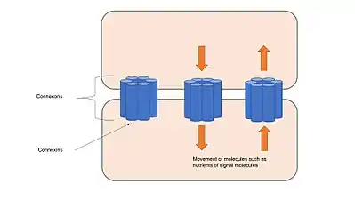
Gap junctions have a very crucial job that cause a tube to form between the 2 cells allowing in the transport of ions and water. Gap junction tubes help the cells to spread electrochemical signals from cell to cell. Electrochemical signals are the product of action potentials that occur in neurons and cardiac cells. Without gap junctions, we would not be able to have a beating heart or functioning nervous system.

Tight signals
Tight signals are just about what the sound like. The 2 cells are squished up against each other, directly connecting the 2 cells membranes, but the contents of the cell are not connected since there is no tube running between the cells. This type of cell connection takes place where certain fluids need to be contained in certain parts of the body like intestines, kidneys, and the bladder. This junction forms a water tight seal, prohibiting the fluids contained in these organs from circulating at will through the body.
Desmosome cell junctions
Desmosome cell junctions physically hold the cells together, but do not allow the cells to pass materials between each other like in gap junction. Desmosome junctions connect the cell with a thread like substance that also connect to the cytoskeleton aiding in the structural support of the cell. These types of junctions are found in areas of the body that undergo a lot of stress, require a lot of flexibility, and movement such as the epidermis and intestines. Desmosomes contain the molecule Cadherins which are also signal receptors. Cadherin of one cell works as the receptor for cadherin in neighbouring cell. Cadherin plays role in contact inhibition [5]
When cells have a communication breakdown
The breakdown of cell communication result in many forms of diseases and the different types of communication breakdown produce different diseases. Multiple sclerosis (MS) is the product when a signal is lost and does not reach its target. In MS the nerve cells protective wrapping found in the brain and spinal cord, get destroyed affecting the nerve cells where they can no longer send signals from one part of the brain to another resulting in the loss of functions such as movement. When the target receptor ignores the signal completely, we end up with the diseases type 1 and type 2 diabetes. In type 1 diabetes the insulin signal is unable to be produced, while in type 2 diabetes the cells have lost the ability to respond to the signals, resulting in abnormally high and dangerous sugar levels in the blood.[6] A stroke results in the production of too much signaling, where dying brain cells release a large amount of glutamate, killing off healthy brain cells leading to widespread damage to the brain. Glutamate is the molecule responsible for many functions in the brain when produced in low concentrates, but when produced in high concentrations is extremely toxic. Excitotoxicity is the spread of highly concentrated glutamine that kill off healthy brain cells not affected by the stroke. Multiple breakdowns in cell communication result in the uncontrolled growth of cells. When one breakdown occurs, the cell gains the ability to grow and divide with out the signal telling it to do so. A cell has the ability to activate a self-destruct sequence (RNAi) to control the unregulated growth of the cell, but when multiple breakdowns occur, the cell loses the ability to self-destruct and the cell divides uncontrollably mutating and creating a tumor. Further cell communication causes blood cells to grow inside the tumor making it grow larger, while more signaling allow the cancerous cells to be spread throughout the body.[7]
RNA interference
In cells, DNA is housed in the nucleus and it never leaves. The DNA within the nucleus is transcribed by tRNA, and the transcriptions of the DNA become RNA. The RNA (ribonucleic acid) leaves the nucleus to float freely around the cytoplasm, and it contains the instructions of the DNA that are vital to the coding, decoding, regulation and expression of genes. The ribosomes then take the RNA messages and turn them into proteins that build cells. When a virus enters a cell and inserts its DNA code into the nucleus, it gets transcribed and released into the cell for the ribosome to make it into a protein. This is how viral infections occur. The cell then explodes with the overproduction of the virus, releasing it into the body to infect any and all other cells it finds. It is theorized that through evolution, cells developed a defense system called RNA interference (RNAi) to stop the production of the proteins that have suspicious mirror image messages of the RNA. They don’t just destroy the suspicious messages, but the correct messages as well to stop all production of that message. This is a cell’s self-destruct mechanism and every cell, plant and animal, have RNAi: a way to turn off the production of a certain gene inside the protein.[8]
RNAi therapy
RNAi therapy is currently being tested in the treatment of cancer. Scientists are trying to harness the RNAi's ability to destroy the genetic code that gets expressed as a cancer cell.[9]
Communication in cancer
Cancer cells will communicate via gap junctions most of the time, and the proteins that form these gap junctions are known as connexins. These connexins have been shown to suppress cancer cells, but this suppression is not the only thing that connexins facilitates. Connexins can also promote tumor progression; therefore, this makes connexins only conditional tumor suppressors.[10] However, this relationship that connects cells makes the spreading of drugs through a system much more effective as small molecules can pass through gap junctions and spread the drug much more quickly and efficiently.[10] The idea that increasing cell communication, or more specifically, connexins, to suppress tumors has been a long, ongoing debate[11] that is supported by the fact that so many types of cancer, including liver cancer, lack the cell communication that characterizes normal cells.
References
- https://www.nature.com/scitable/topic/cell-communication-14122659/
- "The Inside Story of Cell Communication". learn.genetics.utah.edu. Retrieved 2018-11-12.
- "Cell communication I". projects.ncsu.edu. Retrieved 2018-11-12.
- Cell Junctions, retrieved 2018-11-12
- Cell Biology by Pollard et al
- "Big Picture". Big Picture. Retrieved 2018-11-12.
- "When Cell Communication Goes Wrong". learn.genetics.utah.edu. Retrieved 2018-11-12.
- FloatingJetsam (2013-06-27), Nova: RNAi, retrieved 2018-11-12
- Mansoori B, Sandoghchian Shotorbani S, Baradaran B (December 2014). "RNA interference and its role in cancer therapy". Advanced Pharmaceutical Bulletin. 4 (4): 313–21. doi:10.5681/apb.2014.046. PMC 4137419. PMID 25436185.
- Naus CC, Laird DW (June 2010). "Implications and challenges of connexin connections to cancer". Nature Reviews. Cancer. 10 (6): 435–41. doi:10.1038/nrc2841. PMID 20495577.
- Loewenstein WR, Kanno Y (March 1966). "Intercellular communication and the control of tissue growth: lack of communication between cancer cells". Nature. 209 (5029): 1248–9. doi:10.1038/2091248a0. PMID 5956321.
Further reading
- "The Inside Story of Cell Communication". learn.genetics.utah.edu. Retrieved 2018-10-20.
- "When Cell Communication Goes Wrong". learn.genetics.utah.edu. Retrieved 2018-10-24.