Centrocyte
A centrocyte generally refers to a B cell with a cleaved nucleus,[2] as may appear in e.g. follicular lymphoma.[3]
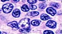
Histopathology of centrocytes in a follicular lymphoma. They have a thick nuclear membrane and prominent nucleoli.[1]
During B cell development, centrocytes are formed following the cessation of centroblast proliferation. Centrocytes test their newly mutated antigen-binding sites (CDR loops) by re-expressing antigen on their surface.
Centrocyte can also refer to a cell with a protoplasm that contains single and double granules of varying size stainable with hematoxylin, as seen in lesions of lichen planus,[2] or a nondividing, activated B cell that expresses membrane immunoglobulin.[2]
References
- Anubha Bajaj (2018). "Susceptive, Supplemented, Stockpiled - Follicular Lymphoma" (PDF). Oncology and Cancer Case Reports. 4 (3).
- " This is an open-access article distributed under the terms of the Creative Commons Attribution License, which permits unrestricted use, distribution, and reproduction in any medium, provided the original author and source are credited." - Stedman's Medical Dictionary. 2006 Archived 2011-09-30 at the Wayback Machine
- Table 12-8 in: Mitchell, Richard Sheppard; Kumar, Vinay; Abbas, Abul K.; Fausto, Nelson. Robbins Basic Pathology. Philadelphia: Saunders. ISBN 1-4160-2973-7. 8th edition.
This article is issued from Wikipedia. The text is licensed under Creative Commons - Attribution - Sharealike. Additional terms may apply for the media files.