FtsK
FtsK is a 1329 amino acid protein involved in bacterial cell division and chromosome segregation.[1] FtsK stands for "Filament temperature sensitive mutant K" because at high temperatures the mutant bacterial cell fails to divide and long filaments develop instead. FtsK, and specifically its C-domain, functions as DNA pump, interacts with other cell division proteins, and intervenes in the regulation of the Xer recombination process. FtsK belongs to the AAA (ATPase Associated with various cellular Activities) superfamily and is present in most bacteria.[2]
| Filament temperature sensitive mutant K | |
|---|---|
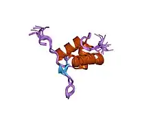 Molecular structure of FtsK C terminal domain. | |
| Identifiers | |
| Symbol | ? |
Structure
FtsK is a transmembrane protein composed of three domains: FtsKN, FtsKL, and FtsKC.[3] Through its N- domain and C-domain FtsK works to coordinate cell division and chromosome differentiation. The FtsKN domain is embedded in the cellular membrane by four transmembrane α-helices.[4] The FtsKL domain extends from the membrane into the cytoplasm.[4] This linking domain varies in length across many bacteria.[4] Found at the cytoplasmic end of the linker domain, the FtsKC segment of the protein is responsible for enabling the activity of the Xer recombination system upon the formation of a chromosome dimer.[4]
Additionally, the FtsKC domain is composed of three subdomains: α, β, and γ.[3] The α and β subunits aggregate to form a hexamer that possesses the ability to translocate DNA through ATP hydrolysis.[3][4] The ATP hydrolysis sites are found on the β subunits of the hexamer.[4] The γ domain is responsible for the control of the hexamer.[4] It mediates the attachment of the hexamer to double-stranded DNA, controls the directionality of the translocase, and initiates chromosome dimer segregation.[4]
Mechanism of Action
The dif Site
The dif site is found at the intersection between the monomers of the chromosome dimer.[4] It corresponds to where chromosomal replication ceased and is also the site of Xer mediated segregation.[3] Translocation of the FtsKC hexamer stops when it reaches the location of the Xer recombinase complex that is associated with the dif site.[3]
Binding Site
Guanosine rich areas of DNA, which are found at the ends of the dif region, are the sites of translocation initiation.[4] These sites are referred to as KOPS motifs.[4] Upon binding a KOPS motif, the FtsK hexamer forms and proceeds towards the dif region.[3][4] Movement toward the dif region is facilitated by the polarity of the KOPS motif.[4]
Translocation
There are three proposed mechanisms of DNA translocation: the rotary inchworm, the staircase, and the revolution mechanism.[4] The rotary inchworm mechanism involves two points of contact between DNA and the subunits of the FtsKC hexamer.[4] These points of contact correspond to an α and a β domain.[4] A conformational change in the α subunit can cause the DNA to shift.[4] This shift is followed by a conformational change in the β subunit (which also causes the DNA to move). The repeated conformational changes lead to the translocation of DNA.[4]
Conversely, the staircase mechanism sees the α and β subunits of the hexamer interacting with the double-stranded DNA in a sequential and overlapping manner.[4] Conformational changes in each subunit cause movement in the spatial position of the DNA strand.[4] Additionally, the revolution mechanism entails the passing of DNA through a channel formed by the hexameric FtsKC domain.[4] In general, the chromosome dimer is translocated so that the site of resolution is near the divisome and so one copy of the genetic material ends up in each daughter cell.[4] FtsK is the fastest DNA translocation pump, with rates of up to 7 kb s−1 it is also a very efficient one.[5]
Recombinase (Xer D) Activation
During bacterial replication, in the presence of a dimer the XerCD mechanism is introduced to divide the dimer into two monomers. FtsK is responsible for the activity of the Xer recombination reaction. Specifically, FtsKc is summoned if a chromosome dimer is present at the mid-cell point.[5] The Xer mechanism is activated by overexpression of FtsK, therefore it appears that FtsK activates the Xer recombination. FtsK turns on the activity of XerCD upon expenditure of ATP.[1]
The recombination apparatus is made up of four monomers, two being Xer D and two being Xer C, that belong to a family of tyrosine recombinases.[3] The interaction of Xer D and the γ subunit of FtsKC results in the activation of the recombinase.[3] Contact between Xer D and the γ subunit is facilitated by the translocation of DNA.[4] Specifically, translocation stops when the FtsKc hexamer reaches the dif site.[4]
Role in Cell Division
FtsK has been shown to be a part of the divisome of bacteria and couple cell division with the resolution of dimers.[4] FtsKN is thought to both stabilize the septum and aid in the recruitment of other proteins to the site of cell division.[4] Recent research has shed light on the role of FtsK in membrane synthesis and has discovered that the L-domain is also important in the building of the septum.[4] There is evidence to suggest that the N terminus is not the only part of FtsK that is involved in cell division. In an experiment done by Dubarry, a suppressor mutation allowed the cells to survive without FtsKN.[6] Parts of FtsK were linked to integral membrane proteins. Subsequently, only those proteins that contained the linker region were able to restore normal cell growth, thus providing convincing evidence that the linker region of FtsK plays an important role in cell division.[6] Other studies have shown that part of the FtsKN domain (which is in the periplasm) is involved in the construction of the cell wall.[4]
Phylogeny
FtsK is part of the AAA motor ATPases. The phylogeny tree of FtsK ties back to the split between ssDNA and dsDNA translocases where TraB, FtsK, T4CPs and VirB4s arise. Each of these show structural similarities and the parent branch of FtsK arose along with other branches of TraB, TcpA, and FtsK. Although FtsK has its own phylogeny specifications and branches within, TraB is similar to a sister protein branch that can trace back to the timeline of FtsK. A common protein that derives from one of the phylogeny branches of FtsK is SpoIIIE which is essential during chromosome segregation. FtsK is found in most bacteria including E. coli , Staphyloccus, and Streptomycetes and in a select amount of Archaea and the phylogenetic tree is similar to that of bacteria. However the difficulty within exact dating of the diversification within its phylogeny is that proteins have variety in their branch lengths which makes it difficult to follow an exact timeline. The phylogeny of FtsK can therefore be compared to the time that protein groups VirB4/VirD4 diversified and slightly earlier than TraB and TcpA as they only occur in Actinobacteria and Firmicutes.[7]
References
- Aussel L, Barre FX, Aroyo M, Stasiak A, Stasiak AZ, Sherratt D (January 2002). "FtsK Is a DNA motor protein that activates chromosome dimer resolution by switching the catalytic state of the XerC and XerD recombinases". Cell. 108 (2): 195–205. doi:10.1016/s0092-8674(02)00624-4. PMID 11832210.
- Pogliano K, Pogliano J, Becker E (December 2003). "Chromosome segregation in Eubacteria". Current Opinion in Microbiology. 6 (6): 586–93. doi:10.1016/j.mib.2003.10.015. PMC 3919143. PMID 14662354.
- Maloy SR, Hughes K, eds. (2013-03-22). Brenner's encyclopedia of genetics (Second ed.). San Diego. ISBN 9780080961569. OCLC 836404630.
- Crozat E, Rousseau P, Fournes F, Cornet F (2014). "The FtsK family of DNA translocases finds the ends of circles". Journal of Molecular Microbiology and Biotechnology. 24 (5–6): 396–408. doi:10.1159/000369213. PMID 25732341.
- Bigot S, Sivanathan V, Possoz C, Barre FX, Cornet F (June 2007). "FtsK, a literate chromosome segregation machine". Molecular Microbiology. 64 (6): 1434–41. doi:10.1111/j.1365-2958.2007.05755.x. PMID 17511809.
- Grainge I (December 2010). "FtsK--a bacterial cell division checkpoint?". Molecular Microbiology. 78 (5): 1055–7. doi:10.1111/j.1365-2958.2010.07411.x. PMID 21155139.
- Guglielmini, J, Rocha E (February 2013). "Evolution of Conjugation and Type IV Secretion Systems". Molecular Biology and Evolution. 30 (2): 315–31. doi:10.1093/molbev/mss221. PMC 3548315. PMID 22977114.