Immune electron microscopy
Immune electron microscopy , sometimes called immunoelectron microscopy is a method used in electron microscopy for diagnosis of viral infections.[1] The technique was first described in the 1940s using tobacco mosaic virus. The technique was not fully exploited until the 1960s, when June Almeida used it to identify viruses including rubella virus and the 1970s when Albert Kapikian discovered noroviruses using the method. [2]
Principle
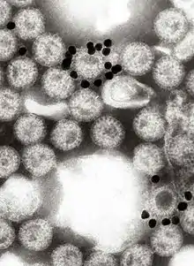
Viruses are suspended, usually in phosphate buffered saline, and antiserum is added. The mixture is warmed, usually to 37°C, centrifuged at 10,000g for a few minutes and the resultant pellet examined by negative stain electron microscopy. Any aggregated virus particles can be identified if the specificity of the antisera is known.[3]
In a technique known as solid phase immune electron microscopy, the antisera is used to coat the electron microscope grid before the virus suspension is added.[4]
References
- Booss 2013, p. 207.
- Booss 2013, p. 209–15.
- Lavazza A, Tittarelli C, Cerioli M. The use of convalescent sera in immune-electron microscopy to detect non-suspected/new viral agents. Viruses. 2015 May 22;7(5):2683-703. doi: 10.3390/v7052683. PMID: 26008707; PMCID: PMC4452926.
- Anderson SR, Parmiter D, Baxa U, Nagashima K. Immunoelectron Microscopy for Visualization of Nanoparticles. Methods Mol Biol. 2018;1682:65-71. doi: 10.1007/978-1-4939-7352-1_7. PMID: 29039094.