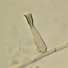
Phialophora verrucosa
Phialophora verrucosa is a pathogenic, dematiaceous fungus that is a common cause of chromoblastomycosis.[1] It has also been reported to cause subcutaneous phaeohyphomycosis and mycetoma in very rare cases.[2] In the natural environment, it can be found in rotting wood,[1] soil,[3] wasp nests,[4] and plant debris.[3] P. verrucosa is sometimes referred to as Phialophora americana, a closely related environmental species which, along with P. verrucosa, is also categorized in the P. carrionii clade.[3]
| Phialophora verrucosa | |
|---|---|
 | |
| Scientific classification | |
| Kingdom: | |
| Division: | |
| Subdivision: | |
| Class: | |
| Order: | |
| Genus: | |
| Species: | P. verrucosa |
| Binomial name | |
| Phialophora verrucosa Medlar (1915) | |
| Synonyms | |
| |
History
The fungus was first isolated by Edgar Mathias Medlar in 1915 from a chronic skin lesion on the buttock of a 22-year-old man in Boston, Massachusetts[5] who presented with verrucous lesions on the buttocks and feet.[6] In consultation with Roland Thaxter, Medlar considered the fungus to represent a previously undescribed genus because the successive separation of the conidia and their maintained attachment to the cup-shaped portion of the sporogenous cells were unique characteristics not seen in any other genus. He named the genus Phialophora, meaning "shallow cup bearer" to represent the characteristic shape and the species epithet verrucosa, in reference to the resemblance of the lesion to "verrucous tuberculosis". Thaxter suggested that P. verrucosa should be classified under the subdivision, 'Chalarae' of Saccardo's classification system.[5]
Morphology and physiology
P. verrucosa produces vase-shaped phialides with dark brown, cup-shaped collarettes.[7] Each phialide is typically 3-4 μm wide and 4-7 μm long.[6] Teardrop-shaped,[3] smooth-walled conidia are formed at the apices of the collarettes and accumulate in clusters. Conidia are typically 2.5 - 4 μm by 1.5 - 3 μm in size.[8] Hyphae are brown, cylindrical, and septate and are composed of thick-walled cells.[5] The hyphae do not produce conidia.[3]P. verrucosa grows well over a range of temperatures, 21–37 °C (70–99 °F) with an optimal growth temperature of 30 °C (86 °F).[9] Colonies grow slowly on oxalic acid and malt extract agar.[3] Grown on Sabouraud's agar at 3 °C (37 °F), the colony attains a diameter of 3–4 cm after 2 weeks incubation.[6]
Ecology
Although P. verrucosa was originally discovered in human tissue, it is known to occur naturally in soil, plant debris,[3] wasp nests,[4] and rotting wood.[1] In a study where multiple strains of P. verrucosa were found growing in rotting wood, soil, and the bark and log of pine trees in Japan, it was found that these isolates from the natural environment had no distinct differences from P. verrucosa isolated from human tissue.[10] P. verrucosa is widespread and can be found in Africa, Asia, Australia, North and South America, and Europe.[11] Most strains of P. verrucosa available in culture collections are derived from human mycoses.[12]
Pathology
P. verrucosa is a common cause of chromoblastomycosis,[1] and a much rarer cause of subcutaneous phaeohyphomycosis and mycetoma.[2] All three diseases have the potential to become chronic.[3] P. verrucosa has also been reported to cause cutaneous infections, prosthetic valve endocarditis, and mycotic keratitis.[13] However, due to its low pathogenicity, P. verrucosa does not often cause infection.[1] Infections caused by P. verrucosa can occur in both immunocompromised individuals, such as individuals who are undergoing immunosuppressive therapies or who have AIDS,[14] as well as in healthy individuals.[1] A healthy individual who became infected with P. verrucosa gained initial exposure through direct contact of the skin with soil containing the fungus.[1] Cases of chromoblastomycosis, subcutaneous phaehyphomycosis, and cutaneous infections caused by P. verrucosa have been reported to present with crusted, warty lesions[15] found on the face,[16] hands,[1] shin,[17] and sole of the foot.[2] Lesions are rarely observed on the back and upper limbs.[18]
Treatment
Antifungal drugs like itraconazole and terbinafine are typically used to treat infections caused by P. verrucosa.[1] Amphotericin B, another antifungal drug, is only used occasionally, as it is cardiotoxic and is unsuitable for long-term therapy.[19] While the spread of chromoblastomycosis to the muscle and bone is usually rare,[15] in cases where antifungal drugs alone are insufficient in controlling the dissemination of the infection, limb amputation is required.[19] Topical heat therapy, such as the use of disposable pocket warmers that sustain a temperature of 40 °C or greater for a period of 12 hours,[1] as well as localized cryotherapy, may be effective in preventing the growth of P. verrucosa and treating lesions.[15] P. verrucosa exhibits some resistance to antifungal drugs, and prescribed treatments often require a combination of antifungal drugs.[20] The use of fluconazole, followed by the combined use of oral itraconazole and the topical application of copper sulphate solution, was reportedly successful in treating a phaehyphomycotic ulcer caused by P. verrucosa.[17] In vitro, different isolates of P. verrucosa respond differently to the same combinations of antifungal drugs. The combination of amphotericin B and terbinafine was observed to cause a synergistic effect for some isolates but cause no effect in others.[20]
References
- Takeuchi, A.; Anzawa, K.; Mochizuki, T.; Takehara, K.; Hamaguchi, Y. (2015). "Chromoblastomycosis caused by Phialophora verrucosa on the hand". European Journal of Dermatology. 25 (3): 274–275. doi:10.1684/ejd.2015.2581. PMID 26066414. S2CID 207262846.
- Turiansky, G. W.; Benson, P. M.; Sperling, L. C.; Sau, P.; Salkin, I. F.; McGinnis, M. R.; James, W. D. (February 1995). "Phialophora verrucosa: A new cause of mycetoma". Journal of the American Academy of Dermatology. 32 (2): 311–315. doi:10.1016/0190-9622(95)90393-3. PMID 7829731.
- Li, Y.; Xiao, J.; de Hoog, G.S.; Wang, X.; Wan, Z.; Yu, J.; Liu, W.; Li, R. (30 June 2017). "Biodiversity and human-pathogenicity of Phialophora verrucosa and relatives in Chaetothyriales". Persoonia. 38 (1): 1–19. doi:10.3767/003158517X692779. PMC 5645179. PMID 29151624.
- Gezuele, E.; Mackinnon, J.E.; Conti-Díaz, I.A. (November 1972). "The frequent isolation of Phialophora verrucosa and Phialophora pedrosoi from natural sources". Sabouraudia. 10 (3): 266–273. doi:10.1080/00362177285190501. PMID 4640043.
- Medlar, E. M. (July 1915). "A New Fungus, Phialophora verrucosa, Pathogenic for Man". Mycologia. 7 (4): 200–203. doi:10.2307/3753363. JSTOR 3753363.
- Kwon-Chung, K.J.; Bennett, John E. (1992). Medical mycology. Philadelphia: Lea & Febiger. p. 338. ISBN 0812114639.
- Liu, ed. by Dongyou (2011). Molecular detection of human fungal pathogens. Boca Raton, Fla.: CRC Press. pp. 346–347. ISBN 9781439812402.CS1 maint: extra text: authors list (link)
- Campbell, Colin K.; Johnson, Elizabeth; Warnock, David W. (2011). Identification of pathogenic fungi (2nd ed.). Chichester: Wiley. ISBN 978-1444330700.
- "CBS 140325". Westerdijk Fungal Biodiversity Institute. Westerdijk Fungal Biodiversity Institute. Retrieved 13 October 2017.
- Iwatsu, T.; Miyaji, M.; Okamoto, S. (1981). "Isolation of Phialophora verrucosa and Fonsecaea pedrosoi from nature in Japan". Mycopathologia. 75 (3): 149–158. doi:10.1007/BF00482809. S2CID 45343939.
- "Phialophora verrucosa". Westerdijk Fungal Biodiversity Institute.
- Untereiner, Wendy A.; Angus, Andrea; Réblová, Martina; Mary-Jane, Orr (June 2008). "Systematics of the Phialophora verrucosa complex: new insights from analyses of β-tubulin, large subunit nuclear rDNA and ITS sequences". Botany. 86 (7): 742–750. doi:10.1139/B08-057.
- Lundstrom, T.S.; Fairfox, M.R.; Dugan, M.C.; Vazquez, J.A.; Chandrasekar, P.H.; Abella, E.; Kasten-Sportes, C. (October 1997). "Phialophora verrucosa infection in a BMT patient". Bone Marrow Transplantation. 20 (9): 789–791. doi:10.1038/sj.bmt.1700969. PMID 9384484.
- Duggan, J.M.; Wolf, M.D.; Kauffiman, C.A. (May 1995). "Phialophora verrucosa infection in an AIDS patient". Mycoses. 38 (5–6): 215–218. doi:10.1111/j.1439-0507.1995.tb00052.x. PMID 8531934. S2CID 22093140.
- Sridhar, K.R. (2009). Frontiers in fungal ecology, diversity and metabolites. New Delhi: I.K. International Pub. House. p. 234. ISBN 9788189866914.
- Hofmann, H; Choi, S. M.; WIlsmann-Theis, D.; Horre, R.; de Hoog, G. S. (2005). "Invasive chromoblastomycosis and sinusitis due to Phialophora verrucosa in a child from Northern Africa". Mycoses. 48 (6): 456–461. doi:10.1111/j.1439-0507.2005.01150.x. PMID 16262887. S2CID 28365592.
- Tendolkar, U.M.; Kerkar, P.; Jerajani, H.; Gogate, A.; Padhye, A.A. (January 1998). "Phaehyphomycotic ulcer caused by Phialophora verrucosa: Successful treatment with itraconazole". Journal of Infection. 36 (1): 122–125. doi:10.1016/S0163-4453(98)93666-0. PMID 9515684.
- Radouane, N.; Hali, F.; Khadir, K.; Soussi, M.; Ouakadi, A.; Marouane, S.; Zamiati, S.; Benchikhi, H. (January 2013). "[Generalized chromomycosis caused by Phialophora verrucosa]". Annales de Dermatologie et de Vénéréologie. 140 (3): 197–201. doi:10.1016/j.annder.2012.10.605. PMID 23466152.
- Queiroz-Telles, Flavio; Esterre, Phillippe; Perez-Blanco, Maigualida; Vitale, Roxana G; Salgado, Claudio Guedes; Bonifaz, Alexandro (February 2009). "Chromoblastomycosis: an overview of clinical manifestations, diagnosis and treatment". Medical Mycology. 47 (1): 3–15. doi:10.1080/13693780802538001. PMID 19085206.
- Li, Y.; Wan, Z.; Li, R. (September 2014). "In Vitro activities of nine antifungal drugs and their combinations against Phialophora verrucosa". Antimicrobial Agents and Chemotherapy. 58 (9): 5609–5612. doi:10.1128/AAC.02875-14. PMC 4135824. PMID 24982078.