Stichocyte
Stichocytes are glandular unicellular cells arranged in a row along the posterior portion of the oesophagus, each of which communicates by a single pore with the lumen of the oesophagus. They contain mitochondria, rough endoplasmic reticulum, abundant Golgi apparatuses, and usually 1 of 2 types of secretory granules, α-granules and β-granules, indicating secretory function [1] [2][3] [4] [5] [6] [7] .[8] Collectively stichocytes form the stichosome. Characteristic of Trichocephalida and Mermithida,[1] two groups of nematodes.
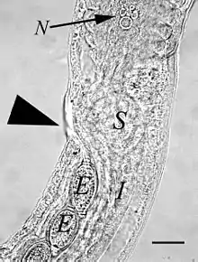
Stichocyte at the posterior extremity of the oesophagus in Capillaria aerophila. N: nucleus of the posteriormost stichocyte. Bar = 50 µm
References
- Chitwood, B. G. & Chitwood, M. B. (1950). Introduction to Nematology (Vol. 1). Baltimore: Monumental Printing Co.doi:10.5962/bhl.title.7355
- Peter J. Gosling. Dictionary of Parasitology. 2005
- Heinz Mehlhorn. Encyclopedia of Parasitology. 3rd Edition 2008
- Larry Roberts, John Janovy. Foundations of Parasitology. 8th edition 2008
- Michael Hutchins, Donna Olendorf. Grzimek's Animal Life Encyclopedia: Lower metazoans and lesser deuterosomes. 2004
- HG Sheffield. Electron microscopy of the bacillary band and stichosome of Trichuris muris and T. vulpis. Journal of Parasitology, 1963
- Despommier, DD; Müller, M (Oct 1976). "The stichosome and its secretion granules in the mature muscle larva of Trichinella spiralis". Journal of Parasitology. 62 (5): 775–85. doi:10.2307/3278960. PMID 978367.
- Lalošević, V.; Lalošević, D.; Capo, I.; Simin, V.; Galfi, A.; Traversa, D. (2013). "High infection rate of zoonotic Eucoleus aerophilus infection in foxes from Serbia". Parasite. 20: 3. doi:10.1051/parasite/2012003. PMC 3718516. PMID 23340229.
This article is issued from Wikipedia. The text is licensed under Creative Commons - Attribution - Sharealike. Additional terms may apply for the media files.