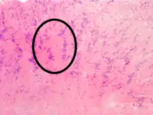
Verocay body
Verocay bodies were first described by Uruguayan neuro-pathologist José Juan Verocay (born: 16 June 1876, Nuevo Paysandú, Uruguay; died: 1927) in 1910. It is a required histopathological finding for diagnosing Schwanommas. Verocay Bodies are a component of Antoni A which are the dense areas of schwannomas located between palisading spindle cells found in neoplasms. Two nuclear palisading regions and an anuclear zone make up 1 Verocay Body.[1]

Naming History: Originally Verocay bodies were called 'neuromas', a term coined by Louis Odier in 1803. The name changed to ‘neuro-fibroma’ under Von Recklinghausen and later in 1935 to ‘neurilemmomas’ curtesy to Arthur Purdy Stout. When Harkin and Reed coined the term 'schwannoma' in 1968, Verocay bodies received its present-day name.[2]
H/P features: 1.Eosinophilic acellular area due to overexpression of lamins.[3]
2.Consisting of reduplicated basement membrane and cytoplasmic processes.[4]
References
- Joshi, Rajiv (2012). "Learning from eponyms: Jose Verocay and Verocay bodies, Antoni A and B areas, Nils Antoni and Schwannomas". Indian Dermatology Online Journal. 3 (3): 215. doi:10.4103/2229-5178.101826. PMID 23189261.
- Wippold FJ, 2nd; Lämmle, M; Anatelli, F; Lennerz, J; Perry, A (November 2006). "Neuropathology for the neuroradiologist: palisades and pseudopalisades". American Journal of Neuroradiology. 27 (10): 2037–41. PMID 17110662.
- Joshi, Rajiv (2012). "Learning from eponyms: Jose Verocay and Verocay bodies, Antoni A and B areas, Nils Antoni and Schwannomas". Indian Dermatology Online Journal. 3 (3): 215. doi:10.4103/2229-5178.101826. PMID 23189261.
- Pytel, Peter; Anthony, Douglas C. (2015). Kumar, Vinay; Abbas, Abul K.; Aster, Jon C. (eds.). Robbins and Cotran Pathologic Basis of Disease. Philadelphia, PA: Elsevier/Saunders. p. 1247. ISBN 978-1-4557-2613-4.