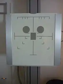
Automatic exposure control
Automatic Exposure Control (AEC) is an X-ray exposure termination device. A medical radiography x-ray exposure is always initiated by a human operator but an AEC detector system may be used to terminate the exposure when a predetermined amount of radiation has been received.[1] The intention of AEC is to provide consistent x-ray image exposure, whether to film, a digital detector or a CT scanner. AEC systems may also automatically set exposure factors such as the X-ray tube current and voltage.[2]

Operation
Projectional Radiography
In projectional radiography an AEC system uses one or more physically thin radiation ionization chambers (the "AEC detector") which is positioned between the patient being x-rayed and the x-ray film cassette. Where low energy x-rays are used such as in mammography the AEC detector is placed behind the image receptor to avoid creating a shadow.[3]:106
In a simple AEC system a weak ionization signal from the AEC detector is integrated as a ramp shaped voltage waveform. This ramp signal rises until it matches a pre-set threshold. At this point the x-ray exposure is terminated.[4] AEC devices are calibrated to ensure that similar exams have linearity in optical density.[5]
Computed Tomography
Modern computed tomography (CT) scanners have AEC systems which aim to maintain image quality for patients of varying sizes, whilst keeping doses as low as reasonably practicable. The systems are also designed to maintain quality with the varying size and attenuation of an individual patient over their length. Implementations vary between manufacturers, some systems are based on a desired noise level in the image, while others are based on a specified reference output (milliampere second, mAs).[6][7]
CT AEC systems use the initial "scanogram", a fixed angle planning view, to determine the relative size of the patient, and variation over their length. The tube output is then adjusted for overall size. The output is also typically modulated for each rotation in response to changes in attenuation over patient length. Some systems adjust output during each rotation, which is known as rotational modulation, based on measured attenuation in the previous rotation.[8]
Advantages
Because patients vary in size and shape, an AEC device is very useful in achieving consistent x-ray film densities, which can be difficult when manually setting exposure factors without AEC.[3]:130
Disadvantages
AEC devices are susceptible to operator error (usually due to mispositioned anatomy or having the incorrect AEC chamber selected).[9] Prosthetic devices such as total hip hardware can also cause the selected ionization chamber to overexpose the image receptor. This is due to the absorption of the x-ray beam into the metal of the hardware as opposed to exposing the ionization chamber.[10]
References
- Sterling, S (1988). "Automatic exposure control: a primer". Radiologic Technology. 59 (5): 421–7. PMID 3290991.
- "Automatic exposure control devices". IAEA Human Health Campus. Retrieved 16 December 2016.
- Dance, D R; Christofides, S; Maidment, A D A; McLean, I D; Ng, K H (2014). Diagnostic radiology physics : a handbook for teachers and students. Vienna: International Atomic Energy Agency. ISBN 978-92-0-131010-1.
- Webb, S (2009). The physics of medical imaging (2nd ed.). London: Taylor & Francis. p. 79. ISBN 9780750305730.
- Doyle, P; Martin, C J (7 November 2006). "Calibrating automatic exposure control devices for digital radiography". Physics in Medicine and Biology. 51 (21): 5475–5485. Bibcode:2006PMB....51.5475D. doi:10.1088/0031-9155/51/21/006. PMID 17047264.
- Söderberg, Marcus; Gunnarsson, Mikael (July 2010). "Automatic exposure control in computed tomography – an evaluation of systems from different manufacturers". Acta Radiologica. 51 (6): 625–634. doi:10.3109/02841851003698206. PMID 20429764. S2CID 9943659.
- Tack, Denis; Kalra, Mannudeep K.; Gevenois, Pierre Alain (2012). Radiation Dose from Multidetector CT. Springer. p. 261. ISBN 9783642245350.
- Keat, Nicholas; ImPACT (2005). "CT scanner automatic exposure control systems". MHRA. Department of Health. Archived from the original (pdf) on 5 December 2017. Retrieved 4 December 2017.
- Walsh, C; Larkin, A; Dennan, S; O'Reilly, G (November 2004). "Exposure variations under error conditions in automatic exposure controlled film–screen projection radiography". The British Journal of Radiology. 77 (923): 931–933. doi:10.1259/bjr/62185486. PMID 15507417.
- Carroll, Quinn B. (2014). Radiography in the digital age: physics, exposure, radiation biology (2nd ed.). Springfield, IL: Charles C Thomas. p. 415. ISBN 9780398080976.
Further reading
- Singh S, Kalra MK, Ali Khawaja RD, Padole A, Pourjabbar S, Lira D, Shepard JA, Digumarthy SR (2014). "Radiation dose optimization and thoracic computed tomography". Radiologic Clinics of North America. 52 (1): 1–15. doi:10.1016/j.rcl.2013.08.004. PMID 24267707.
