Cellular compartment
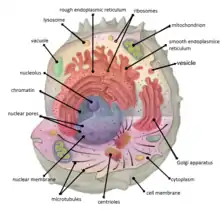
Cellular compartments in cell biology comprise all of the closed parts within the cytosol of a eukaryotic cell, usually surrounded by a single or double lipid layer membrane. These compartments are often, but not always, defined as membrane enclosed regions. The formation of cellular compartments is called compartmentalization.

Both organelles, the mitochondria and chloroplasts (in photosynthetic organisms), are compartments that are believed to be of endosymbiotic origin. Other compartments such as peroxisomes, lysosomes, the endoplasmic reticulum, the cell nucleus or the Golgi apparatus are not of endosymbiotic origin. Smaller elements like vesicles, and sometimes even microtubules can also be counted as compartments.
It was thought that compartmentalization is not found in prokaryotic cells.,[1] but the discovery of carboxysomes and many other metabolosomes revealed that prokaryotic cells are capable of making compartmentalized structures, albeit these are in most cases not surrounded by a lipid bilayer, but of pure proteinaceous built.[2][3][4]
Types
In general there are 4 main cellular compartments, they are:
- The nuclear compartment comprising the nucleus
- The intercisternal space which comprises the space between the membranes of the endoplasmic reticulum (which is continuous with the nuclear envelope)
- Organelles (the mitochondrion in all eukaryotes and the plastid in phototrophic eukaryotes)
- The cytosol
Function
Compartments have three main roles. One is to establish physical boundaries for biological processes that enables the cell to carry out different metabolic activities at the same time. This may include keeping certain biomolecules within a region, or keeping other molecules outside. Within the membrane-bound compartments, different intracellular pH, different enzyme systems, and other differences are isolated from other organelles and cytosol. With mitochondria, the cytosol has an oxidizing environment which converts NADH to NAD+. With these cases, the compartmentalization is physical.
Another is to generate a specific micro-environment to spatially or temporally regulate a biological process. As an example, a yeast vacuole is normally acidified by proton transporters on the membrane.
A third role is to establish specific locations or cellular addresses for which processes should occur. For example, a transcription factor may be directed to a nucleus, where it can promote transcription of certain genes. In terms of protein synthesis, the necessary organelles are relatively near one another. The nucleolus within the nuclear envelope is the location of ribosome synthesis. The destination of synthesized ribosomes for protein translation is rough endoplasmic reticulum (rough ER), which is connected to and shares the same membrane with the nucleus. The Golgi body is also near the rough ER for packaging and redistributing. Likewise, intracellular compartmentalization allows specific sites of related eukaryotic cell functions isolated from other processes and therefore efficient.
Establishment
Often, cellular compartments are defined by membrane enclosure. These membranes provide physical barriers to biomolecules. Transport across these barriers is often controlled in order to maintain the optimal concentration of biomolecules within and outside of the compartment.
References
- Campbell, Neil A.; Reece, Jane B.; Urry, Lisa A.; Cain, Michael L.; Wasserman, Steven A.; Minorsky, Peter V.; Jackson, Robert B. (2008). Biology (8th ed.). p. 559. ISBN 978-0-8053-6844-4.
- Grant, CR; Wan, J; Komeili, A (6 October 2018). "Organelle Formation in Bacteria and Archaea". Annual Review of Cell and Developmental Biology. 34: 217–238. doi:10.1146/annurev-cellbio-100616-060908. PMID 30113887.
- Diekmann, Y; Pereira-Leal, JB (15 January 2013). "Evolution of intracellular compartmentalization". The Biochemical Journal. 449 (2): 319–31. doi:10.1042/BJ20120957. PMID 23240612.
- Cornejo, E; Abreu, N; Komeili, A (February 2014). "Compartmentalization and organelle formation in bacteria". Current Opinion in Cell Biology. 26: 132–8. doi:10.1016/j.ceb.2013.12.007. PMC 4318566. PMID 24440431.