Exophiala phaeomuriformis
Exophiala phaeomuriformis is thermophilic fungus belonging to the genus Exophiala and the family Herpotrichiellaceae.[1][2] it is a member of the group of fungi known as black yeasts, and is typically found in hot and humid locations, such as saunas, bathrooms, and dishwashers.[3] This species can cause skin infections[4] and is typically classified as a Biosafety Risk Group 2 agent.[5]
| Exophiala phaeomuriformis | |
|---|---|
 | |
| Scientific classification | |
| Kingdom: | |
| Division: | |
| Class: | |
| Order: | |
| Family: | |
| Genus: | |
| Species: | E. phaeomuriformis |
| Binomial name | |
| Exophiala phaeomuriformis Matos (2003) | |
| Synonyms | |
| |
History
Exophiala phaeomuriformis is a member of the genus Exophiala, described in 1952 based on E. jeanselmei.[1] Thirty species of Exophiala are currently recognized[6] amongst which Exophiala (Wangiella) dermatitidis is the most common.[7] When studying samples of E. dermatitidis, Takashi Matsumoto and colleagues observed strains with a granular colonial form and distinctive microscopic morphology.[8] Based on the resemblance of these strains to the genus Sarcinomyces, they proposed the new name, S. phaeomuriformis.[8] This taxon was transferred to the genus Exophiala by Tiago Matos and co-workers in 2003 because of its yeast-like morphology (rather than the meristematic form characteristic to members of the genus Sarcinomyces), and its closer DNA homology to the genus Exophiala.[9]
Exophiala phaeomuriformis is a dematiceous (darkly pigmented) fungus and member of the group of fungi known as the black yeasts.[10][11] Black yeasts are an unrelated category of fungi that share yeast-like morphology and possess darkly melanized cell walls.[5] Although their DNA sequences are distinctive, E. phaeomuriformis and E. dermatitidis are so closely related that the two cannot be reliably differentiated morphologically or physiologically.[5][12] Antigenic cross-reactivity suggests that E. phaeomuriformis may have originated as multicellular variant of E. dematitidis.[10]
Growth and morphology
Like many other black yeasts, Exophiala phaeomuriformis is known only by its asexual form and no sexual form has been found.[4][5][8] It is a thermophilic fungus preferring temperatures between 37–42 °C (99–108 °F)[2] but growing at any temperature between 15–42 °C (59–108 °F).[3] Exophilala phaeomuriformis is more sensitive than other black yeasts to salt, incapable of growth at concentrations of sodium chloride exceeding 17%.[3] Like other members of the genus Exophiala, it is able to tolerate a wide range of pH (2.5–12.5).[3]
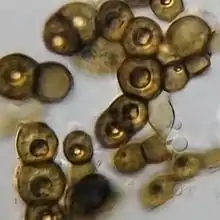
Colonies of E. phaeomuriformis are hyaline, mycoid, and smooth when young[9] but become black, dry, crumbly, raised, and mulberry-like in texture with age.[4][8] Some strains fail to undergo this morphological switch and remain yeast-like in age.[5][8] By contrast, many strains of E. dermatitidis become mycelial with age.[8] Hyphal growth has not been observed in E. phaeomuriformis.[8] Instead, colonies develop from loosely packed, single, and rounded budding yeast cells that are either scattered or aggregated in groups.[5][8] Vegetative cells can either by unicellular or muriform (septate in all planes) or become divided by transverse septa only.[4][8] Yeast cells are thick-walled and spherical or near-spherical in shape.[4][5] Budding cells can have broad bases, occur in chains, and be multilateral, budding in different directions.[9]
Physiology
Like other member of the genus Exophiala, E. phaeomuriformis is saprotrophic, obtaining its energy exclusively from non-living organic materials.[6] When inoculated on a suitable growth medium under optimal conditions, the growth of E. phaeomuriformis is initiated in roughly 3 days;[3] however, when subject to competition, the cells may remain in a stationary state for many weeks prior to the development of visible growth.[3] Similar to E. dematitidis, E. phaeomuriformis is unable to assimilate nitrate, nitrite and melibiose; however it differs in it that some strains are unable to metabolize D-gluconate, D-glucuronate, D-galacturonate and glucono-δ-lactone.[10]
Habitat and ecology
Exophiala phaeomuriformis has a proclivity for environments rich in mono- and polyaromatic compounds, such as hydrocarbons, where it uses these compounds as sources of energy.[3] The species is plurivorous, occurring on a wide range of materials from contaminated soils and toluene rich environments to wild berries and animal feces.[3] It is also found in environments containing the preservative creosote, such as railroad ties where it is an important agent of biodeterioration.[3][13] In indoor environments, E. phaeomuriformis occurs in warm and moist environments such as toilets, saunas, or dishwashers.[2] This species is found world-wide.[14]
Human disease
Exophiala phaeomuriformis is a rare causative agent of phaeohyphomycosis[15] in cutaneous, subcutaneous and deep tissues,[4] and is responsible for 6.4% of infections caused by black yeasts.[7] Infection usually occurs following skin abrasion or penetrating injuries.[11] Exophiala phaeomuriformis can also cause corneal infection following eye exposure to contaminated water.[1] People with cystic fibrosis (CF) are considered abnormally susceptible to Exophiala infections, including E. phaeomuriformis.[14][16] It has been suggested that differences in the microbiota profiles of CF patients may be responsible for this predisposition.[17] Treatment of E. phaeomuriformis involves a combination of surgical debridement and antifungal therapy.[15] A range of antifungal agents including caspofungin, voriconazole, itraconazole, posaconazole, and amphotericin B are active against this species.[16][18] Due to its pathogenic potential, E. phaeomuriformis is regarded as a Biosafety Risk Group 2 agent in the laboratory.[5]
References
- Aggarwal, Shruti; Yamaguchi, Takefumi; Dana, Reza; Hamrah, Pedram (October 2015). "Exophiala phaeomuriformis Fungal Keratitis". Eye & Contact Lens: Science & Clinical Practice. 43 (2): e4–e6. doi:10.1097/ICL.0000000000000193. PMID 26513718.
- Ozhak-Baysan, B.; O unc, D.; Do en, A.; Ilkit, M.; de Hoog, G. S. (6 April 2015). "MALDI-TOF MS-based identification of black yeasts of the genus Exophiala". Medical Mycology. 53 (4): 347–352. doi:10.1093/mmy/myu093. PMID 25851261.
- Döğen, Aylin; Kaplan, Engin; Ilkit, Macit; de Hoog, G. Sybren (3 November 2012). "Massive Contamination of Exophiala dermatitidis and E. phaeomuriformis in Railway Stations in Subtropical Turkey". Mycopathologia. 175 (5–6): 381–386. doi:10.1007/s11046-012-9594-z. PMID 23124309. S2CID 16726797.
- Howard, ed. by Dexter H. (2003). Pathogenic fungi in humans and animals (2. ed.). New York [u.a.]: Dekker. ISBN 0-8247-0683-8.CS1 maint: extra text: authors list (link)
- de Hoog, G.S.; Guarro, J; Gene, J; Figueras, M.J. (1995). Atlas of Clinical Fungi (2nd ed.). Amer Society for Microbiology.
- Woo, Patrick C. Y.; Ngan, Antonio H. Y.; Tsang, Chris C. C.; Ling, Ian W. H.; Chan, Jasper F. W.; Leung, Shui-Yee; Yuen, Kwok-Yung; Lau, Susanna K. P. (January 2013). "Clinical Spectrum of Exophiala Infections and a Novel Exophiala Species, Exophiala hongkongensis". Journal of Clinical Microbiology. 51 (1): 260–267. doi:10.1128/JCM.02336-12. PMC 3536265. PMID 23152554.
- Zeng, J. S.; Sutton, D. A.; Fothergill, A. W.; Rinaldi, M. G.; Harrak, M. J.; de Hoog, G. S. (27 June 2007). "Spectrum of Clinically Relevant Exophiala Species in the United States". Journal of Clinical Microbiology. 45 (11): 3713–3720. doi:10.1128/JCM.02012-06. PMC 2168524. PMID 17596364.
- Matsumoto, T.; Padhye, A.A.; Ajello, L.; McGinnis, M.R. (January 1986). "a new dematiaceous hyphomycete". Medical Mycology. 24 (5): 395–400. doi:10.1080/02681218680000601. PMID 3783362.
- Matos, T.; Haase, G; Gerrits van den Ende, A.H.G; deHoog, G (2003). "Molecular diversity of oligotrophic and neurotropic members of the black yeast genus Exophiala, with accent on E. dermatitidis". Antonie van Leeuwenhoek. 83 (4): 293–303. doi:10.1023/A:1023373329502. PMID 12777065. S2CID 4849346.
- Uijthof; Van Belkum; De Hoog; Haase (25 July 2008). "Exophiala dermatitidis and Sarcinomyces phaeomuriformis: ITS1-sequencing and nutritional physiology". Medical Mycology. 36 (3): 143–151. doi:10.1111/j.1365-280X.1998.00143.x. PMID 9776827.
- Rogers, Everett Smith Beneke, Alvin Lee (1996). Medical mycology and human mycoses. Belmont, Calif.: Star Pub. Co. ISBN 0-89863-175-0.
- Haase, G.; Sonntag, L.; van de Peer, Y.; Uijthof, J. M. J.; Podbielski, A.; Melzer-Krick, B. (March 1995). "Phylogenetic analysis of ten black yeast species using nuclear small subunit rRNA gene sequences". Antonie van Leeuwenhoek. 68 (1): 19–33. doi:10.1007/BF00873289. PMID 8526477. S2CID 2819521.
- K June; Wang (1990). Identification manual for fungi from utility poles in the eastern United States. Rockville, Md.: American Type Culture Collection. pp. 356. ISBN 9780930009311.
- Döğen, Aylin; Ilkit, Macit; de Hoog, G. Sybren (October 2013). "Black yeast habitat choices and species spectrum on high altitude creosote-treated railway ties". Fungal Biology. 117 (10): 692–696. doi:10.1016/j.funbio.2013.07.006. PMID 24119407.
- Kwon-Chung, K.J.; Bennett, John E. (1992). Medical mycology. Philadelphia: Lea & Febiger. ISBN 0-8121-1463-9.
- Packeu, A.; Lebecque, P.; Rodriguez-Villalobos, H.; Boeras, A.; Hendrickx, M.; Bouchara, J.-P.; Symoens, F. (11 May 2012). "Molecular typing and antifungal susceptibility of Exophiala isolates from patients with cystic fibrosis". Journal of Medical Microbiology. 61 (Pt_9): 1226–1233. doi:10.1099/jmm.0.042317-0. PMID 22580912.
- Lebecque, Patrick; Leonard, Anissa; Huang, Daniel; Reychler, Grégory; Boeras, Anca; Leal, Teresinha; Symoens, Françoise (November 2010). "Exophiala (Wangiella) dermatitidis and cystic fibrosis – Prevalence and risk factors". Medical Mycology. 48 (O1): S4–S9. doi:10.3109/13693786.2010.495731. PMID 21067329.
- Rivard, Robert G.; McCall, Suzanne; Griffith, Matthew E.; Hawley, Joshua S.; Ressner, Roseanne A.; Borra, Himabindu; Moon, James E.; Beckius, Miriam L.; Murray, Clinton K.; Hospenthal, Duane R. (January 2007). "Efficacy of caspofungin and posaconazole in a murine model of disseminated infection". Medical Mycology. 45 (8): 685–689. doi:10.1080/13693780701390157. PMID 17885951.