Fat necrosis
Fat necrosis is a form of necrosis characterized by the action upon fat by digestive enzymes.[1]
| Fat necrosis | |
|---|---|
| Other names | also known as Balser's necrosis |
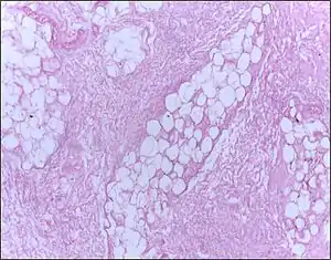 | |
| Micrograph of breast tissue showing fat necrosis. H&E stain | |
| Specialty | Pathology |
In fat necrosis the enzyme lipase releases fatty acids from triglycerides. The fatty acids then complex with calcium to form soaps. These soaps appear as white chalky deposits.[2]
It is usually associated with trauma of the pancreas or acute pancreatitis.[2][3] It can also occur in the breast,[4] the salivary glands[5] and neonates after a traumatic delivery.[6]
Signs and symptoms
Signs and Symptoms of fat necrosis are presented below
- masses that seem to be irregular beneath the skin, by touch these should feel smooth and rounded.
- tenderness in areas with masses
- skin tethering
- dimpling
- nipple retraction[7]
Causes
Fat necrosis, although most commonly found in the breast, can also be found in various parts of the body and is caused by severe trauma.Some example of severe trauma to the breast include but are not limited to:
- radiotherapy
- cyst aspiration
- biopsy
- lumpectomy
- reduction mammoplasty
Fat necrosis can also be caused by polyarteritis nodosa or weber-Christer disease and other times causes may be unknown and can occur randomly.[8]
Mechanism/pathophysiology
Fat necrosis is inflammation that is free from contaminants (bacteria or viruses) saponification takes place, this is the process of fat converting to soap through the use of heat. Lipase is the enzyme used to conduct this conversion, it forms a complex with calcium to form this process.
When fat necrosis first occurs due to bleeding in fat, this area can become firm. Next, the process mentioned above takes place saponification and this causes the lesion to become discolored (yellow), it continues to then calcify which turns it into a white color, and in the last step of this process, fibrosis occurs causing a grayish-yellow mass.
After this process is complete a scar may form due to the inflammation and can result in fibrosis, which can surround dissipated fats or oils. Cystic degeneration may also occur if adipose cells contents are released, the lining of cysts may also calcify.[9]
Fat necrosis has also been associated with various types of pancreatitis, such as carcinoma of the pancreas and pancreatic trauma. Pancreatic lipase and colipase, are the agents said to cause this is, they are said to escape the pancreas while the disease is developing. The mechanism of these factors is caused due to the attack of adipose tissue, which leads to the production of foci of fat necrosis, however, the precise mechanism is unknown. The pathology of fat necrosis is not confined to the peritoneal-retroperitoneal region, however direct contact with the factors presented is the major cause. In other various patients, fat necrosis will often involve the peripheral tissues, particularly in subcutaneous adipose tissue in all regions of the body, joints of feet and hands, and within the bone marrow. Individuals who suffer from primary pancreatic disease polyarthritis, osteolytic defects, and skin lesions are exhibited on sites that are involved.[10]
Diagnosis
Although fat necrosis can be diagnosed through a routine checkup with a physician, called a physical. A patient can also perform a physical checkup on themselves. For additional diagnosis, a physician would request one or more of the following scans or tests.
- CT scan
- Mammography test
- Sonography test
- MRI test[8]
Prevention/treatment
When fat necrosis is being felt by a physician or patient, it may feel larger, smaller, unchanged, or not felt at all (resolved). Fat necrosis usually does not require surgery, it usually requires a patient to meet with their physician and as long as the pain is not present there is nothing to be concerned about unless the patient is concerned about cosmetic abnormalities. However, if pain is present surgery is a form of treatment a patient can consider.
To keep track of benign fat necrosis a yearly mammogram is taken in order to observe it.
However, if fat necrosis consists of oily fluid a physician will go in with a needle to remove this liquid, which may be causing discomfort. Excision may be needed if the mass becomes solid or causes a cosmetic abnormality.
Prognosis
Although getting diagnosed with fat necrosis of any kind can be a great cause for concern, as most individuals may mistake it for a malignant tumor. Fat necrosis of the breast is a prognosis that is benign and does not increase an individual's risk for various cancers. An individual's life expectancy does not decrease with this diagnosis.
Epidemiology
Fat necrosis in the breast occurs around 0.6%, this represents 2.75% of lesions that end up being benign. However, 0.8% of fat necrosis occurs from tumors of the breast, 1% - 9% occurs in breast reduction surgery. Individuals that are high risk include women around the age of 50yrs along with pendulous breasts.[6]
References
- "Cell Injury".
- Kumar, Vinay; Abbas, Abul K.; Fausto, Nelson; & Mitchell, Richard N. (2007). Robbins Basic Pathology (8th ed.). Saunders Elsevier. pp. 10-11 ISBN 978-1-4160-2973-1
- "fat necrosis" at Dorland's Medical Dictionary
- Lövey K, Fodor J, Major T, et al. (November 2007). "Fat necrosis after partial-breast irradiation with brachytherapy or electron irradiation versus standard whole-breast radiotherapy--4-year results of a randomized trial". Int. J. Radiat. Oncol. Biol. Phys. 69 (3): 724–31. doi:10.1016/j.ijrobp.2007.03.055. PMID 17524571.
- "Archived copy". Archived from the original on 2011-04-14. Retrieved 2011-09-19.CS1 maint: archived copy as title (link)
- Rice, Hannah; Warland, Jane (October 2013). "Bearing Witness: Midwives experiences of witnessing traumatic birth". Women and Birth. 26: S39. doi:10.1016/j.wombi.2013.08.215. ISSN 1871-5192.
- Vasei, Narges; Shishegar, Azita; Ghalkhani, Forouzan; Darvishi, Mohammad (2019-06-11). "Fat necrosis in the Breast: A systematic review of clinical". Lipids in Health and Disease. 18. doi:10.1186/s12944-019-1078-4. ISSN 1476-511X. PMC 6560815. PMID 31185981.
- Kerridge, William D.; Kryvenko, Oleksandr N.; Thompson, Afua; Shah, Biren A. (2015-03-16). "Fat Necrosis of the Breast: A Pictorial Review of the Mammographic, Ultrasound, CT, and MRI Findings with Histopathologic Correlation". Radiology Research and Practice. Retrieved 2020-11-12.
- Genova, Rafaella; Garza, Robert F. (2020), "Breast Fat Necrosis", StatPearls, Treasure Island (FL): StatPearls Publishing, PMID 31194348, retrieved 2020-11-12
- Lee, P. C.; Howard, J. M. (May 1979). "Fat necrosis". Surgery, Gynecology & Obstetrics. 148 (5): 785–789. ISSN 0039-6087. PMID 432796.