Fuchs spot
The Fuchs spot (also known as Förster-Fuchs' Spot[1]), is a degeneration of the macula in case of high myopia. It is named after the two persons who first described it: Ernst Fuchs, who described a pigmented lesion in 1901, and Forster, who described subretinal neovascularisation in 1862.[2] It occur due to proliferation of retinal pigment epithelium associated with choroidal hemorrhage.[1] The size of the spots are proportionate to the severity of the pathological myopia.
| Fuchs spot | |
|---|---|
| Other names | Forster-Fuchs' retinal spot |
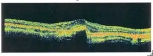 | |
| An Optical coherence tomography (OCT) of the retina, showing a Fuchs spot | |
Symptoms
First signs of a Fuchs spot are distorted sight of straight lines near the fovea, which some days later turn to the typical well-circumscribed patches after absorption of haemorrhage, and a pigmented scar remains. As in macular degeneration, central sight is affected. Atrophy leads to the loss of two or more lines of the Snellen chart.
Treatment
Fuchs spots are caused by regression of choroidal neovascularization.[3] Since it is a medical sign, treatment is given for the actual cause. Photothermal laser ablation, photodynamic therapy, anti-VEGF therapy, or a combination of these are the treatment options of choroidal neaovascularization due to pathological myopia.[3][1]
See also
References
- Kumar, Atul; Chawla, Rohan; Kumawat, Devesh; Pillay, Ganesh (2017). "Insight into high myopia and the macula". Indian Journal of Ophthalmology. 65 (2): 85–91. doi:10.4103/ijo.IJO_863_16. ISSN 0301-4738.
- "Forster-Fuchs' Retinal Spot". patient.info. Retrieved 24 December 2012.
- "Pathologic myopia (myopic degeneration) - EyeWiki". eyewiki.aao.org.