Glycoside hydrolase family 67
In molecular biology, glycoside hydrolase family 67 is a family of glycoside hydrolases.
| Glycosyl hydrolase family 67 N-terminus | |||||||||
|---|---|---|---|---|---|---|---|---|---|
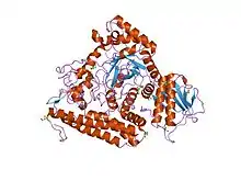
 the 1.7 a crystal structure of alpha-d-glucuronidase, a family-67 glycoside hydrolase from bacillus stearothermophilus t-1 | |||||||||
| Identifiers | |||||||||
| Symbol | Glyco_hydro_67N | ||||||||
| Pfam | PF03648 | ||||||||
| InterPro | IPR005154 | ||||||||
| SCOP2 | 1h41 / SCOPe / SUPFAM | ||||||||
| CAZy | GH67 | ||||||||
| |||||||||
| Glycosyl hydrolase family 67 middle domain | |||||||||
|---|---|---|---|---|---|---|---|---|---|
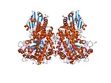
 pseudomonas cellulosa e292a alpha-d-glucuronidase mutant complexed with aldotriuronic acid | |||||||||
| Identifiers | |||||||||
| Symbol | Glyco_hydro_67M | ||||||||
| Pfam | PF07488 | ||||||||
| InterPro | IPR011100 | ||||||||
| SCOP2 | 1h41 / SCOPe / SUPFAM | ||||||||
| CAZy | GH67 | ||||||||
| |||||||||
| Glycosyl hydrolase family 67 C-terminus | |||||||||
|---|---|---|---|---|---|---|---|---|---|
 the 1.7 a crystal structure of alpha-d-glucuronidase, a family-67 glycoside hydrolase from bacillus stearothermophilus t-1 | |||||||||
| Identifiers | |||||||||
| Symbol | Glyco_hydro_67C | ||||||||
| Pfam | PF07477 | ||||||||
| InterPro | IPR011099 | ||||||||
| SCOP2 | 1h41 / SCOPe / SUPFAM | ||||||||
| CAZy | GH67 | ||||||||
| |||||||||
Glycoside hydrolases EC 3.2.1. are a widespread group of enzymes that hydrolyse the glycosidic bond between two or more carbohydrates, or between a carbohydrate and a non-carbohydrate moiety. A classification system for glycoside hydrolases, based on sequence similarity, has led to the definition of >100 different families.[1][2][3] This classification is available on the CAZy web site,[4][5] and also discussed at CAZypedia, an online encyclopedia of carbohydrate active enzymes.[6][7]
Glycoside hydrolase family 67 includes alpha-glucuronidases, these are components of an ensemble of enzymes central to the recycling of photosynthetic biomass, remove the alpha-1,2 linked 4-O-methyl glucuronic acid from xylans.
Members of this family consist of three structural domains. Deletion mutants of alpha-glucuronidase from Bacillus stearothermophilus have indicated that the central region is responsible for the catalytic activity. Within this central domain, the invariant Glu and Asp (residues 391 and 364 respectively from Bacillus stearothermophilus) are thought to form the catalytic centre.[8] The C-terminal region of alpha-glucuronidase is mainly alpha-helical. It wraps around the catalytic domain, making additional interactions both with the N-terminal domain of its parent monomer and also forming the majority of the dimer-surface with the equivalent C-terminal domain of the other monomer of the dimer.[9]
References
- Henrissat B, Callebaut I, Fabrega S, Lehn P, Mornon JP, Davies G (July 1995). "Conserved catalytic machinery and the prediction of a common fold for several families of glycosyl hydrolases". Proceedings of the National Academy of Sciences of the United States of America. 92 (15): 7090–4. Bibcode:1995PNAS...92.7090H. doi:10.1073/pnas.92.15.7090. PMC 41477. PMID 7624375.
- Davies G, Henrissat B (September 1995). "Structures and mechanisms of glycosyl hydrolases". Structure. 3 (9): 853–9. doi:10.1016/S0969-2126(01)00220-9. PMID 8535779.
- Henrissat B, Bairoch A (June 1996). "Updating the sequence-based classification of glycosyl hydrolases". The Biochemical Journal. 316 ( Pt 2) (Pt 2): 695–6. doi:10.1042/bj3160695. PMC 1217404. PMID 8687420.
- "Home". CAZy.org. Retrieved 2018-03-06.
- Lombard V, Golaconda Ramulu H, Drula E, Coutinho PM, Henrissat B (January 2014). "The carbohydrate-active enzymes database (CAZy) in 2013". Nucleic Acids Research. 42 (Database issue): D490–5. doi:10.1093/nar/gkt1178. PMC 3965031. PMID 24270786.
- "Glycoside Hydrolase Family 67". CAZypedia.org. Retrieved 2018-03-06.
- CAZypedia Consortium (December 2018). "Ten years of CAZypedia: a living encyclopedia of carbohydrate-active enzymes" (PDF). Glycobiology. 28 (1): 3–8. doi:10.1093/glycob/cwx089. hdl:21.11116/0000-0003-B7EB-6. PMID 29040563.
- Shoham Y, Zaide G, Shallom D, Shulami S, Zolotnitsky G, Golan G, Baasov T, Shoham G (2001). "Biochemical characterization and identification of catalytic residues in alpha-glucuronidase from Bacillus stearothermophilus T-6". Eur. J. Biochem. 268 (10): 3006–3016. doi:10.1046/j.1432-1327.2001.02193.x. PMID 11358519.
- Nurizzo D, Nagy T, Gilbert HJ, Davies GJ (April 2002). "The structural basis for catalysis and specificity of the Pseudomonas cellulosa alpha-glucuronidase, GlcA67A". Structure. 10 (4): 547–56. doi:10.1016/s0969-2126(02)00742-6. PMID 11937059.