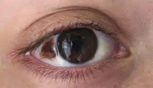
Nevus of Ota
Nevus of Ota is a blue hyperpigmentation[3] that occurs on the face, most often appearing on the white of the eye. It also occurs on the forehead, nose, cheek, periorbital region, and temple.[4]
| Nevus of Ota | |
|---|---|
| Other names | Congenital melanosis bulbi,[1] nevus fuscoceruleus ophthalmomaxillaris, oculodermal melanocytosis,[2]:700 oculomucodermal melanocytosis[1] |
 | |
| Specialty | Oncology |
Cause
Nevus of Ota is caused by the entrapment of melanocytes in the upper third of the dermis. It is found only on the face, most commonly unilaterally, rarely bilaterally and involves the first two branches of the trigeminal nerve. The sclera is involved in two-thirds of cases (causing an increased risk of glaucoma). It should not be confused with Mongolian spot, which is a birthmark caused by entrapment of melanocytes in the dermis but is located in the lumbosacral region. Women are nearly five times more likely to be affected than men, and it is rare among Caucasian people.[6] Nevus of Ota may not be congenital, and may appear during puberty.
Skin treatment
A Q-switched 1064 nm laser has been successfully used to treat the condition.[7][8] The Q-Switched Lasers (694 nm Ruby, 755 nm Alexandrite or 1064 nm Nd-YAG) with their high peak power and pulse width in nano second range are best suited to treat various epidermal, junctional, mixed and dermal lesions The Q-switched 1064 nm Nd-YAG is an ideal choice to treat dermal pigment as in nevus of Ota and in darker skin types, as it reduces the risk of epidermal injury and pigmentary alterations. The pigment clearance may be expected to be near total. Usually a number of treatment sessions with an interval of minimum six weeks are required to achieve total or near total clearance. The number of treatments depend mainly on the severity of the lesion. Darker the lesion, more are the treatments required. It also depends to some extent on the quality of the laser system used and its energy output. Last but not the least, experience of the laser surgeon plays a significant role in achieving early and good clearance.[9]
Treatment
A specific form of conjunctivoplasty may help somewhat.
Notable cases
- Actress Daniela Ruah[6] of NCIS: Los Angeles, right eye
- Actor Eriq La Salle of ER, left eye
See also
- Nevus of Ito
- List of cutaneous conditions
References
- Rapini, Ronald P.; Bolognia, Jean L.; Jorizzo, Joseph L. (2007). Dermatology: 2-Volume Set. St. Louis: Mosby. pp. 1720–22. ISBN 978-1-4160-2999-1.
- James, William D.; Berger, Timothy G.; et al. (2006). Andrews' Diseases of the Skin: Clinical Dermatology. Saunders Elsevier. ISBN 0-7216-2921-0.
- Chan HH, Kono T (2003). "Nevus of Ota: clinical aspects and management". Skinmed. 2 (2): 89–96, quiz 97–8. doi:10.1111/j.1540-9740.2003.01706.x. PMID 14673306. Archived from the original on 2019-12-16. Retrieved 2009-02-28.
- Mohan, R. P. S.; Verma, S.; Singh, A. K.; Singh, U. (2013). "'Nevi of Ota: the unusual birthmarks': a case review". BMJ Case Reports. 2013: bcr2013008648. doi:10.1136/bcr-2013-008648. PMC 3618781. PMID 23456162.
- "eMedicine - Nevi of Ota and Ito : Article by Harvey Lui". Retrieved 2008-03-22.
- "Daniela Ruah Officially Checks In". Esquire. September 2011. Retrieved 9 June 2016.
- Geronemus, RG (Dec 1992). "Q-switched ruby laser therapy of nevus of Ota". Archives of Dermatology. 128 (12): 1618–22. Bibcode:1992SPIE.1643..284G. doi:10.1001/archderm.1992.04530010056008. PMID 1456756.
- Watanabe S, Takahashi H (1994). "Treatment of nevus of Ota with the Q-switched ruby laser". New England Journal of Medicine. 331 (26): 1745–50. doi:10.1056/NEJM199412293312604. PMID 7984195.
- Uddhav A. Patil, Lakshyajit D. Dhami. Overview of Lasers: Indian Journal of Plastic Surgery Supplement 2008 Vol 41 S101-113.
| Classification | |
|---|---|
| External resources |
