TMEM126A
Transmembrane protein 126A is a mitochondrial transmembrane protein of unknown function coded for by the TMEM126A gene.[5][6]
| TMEM126A | |||||||||||||||||||||||||
|---|---|---|---|---|---|---|---|---|---|---|---|---|---|---|---|---|---|---|---|---|---|---|---|---|---|
| Identifiers | |||||||||||||||||||||||||
| Aliases | TMEM126A, OPA7, transmembrane protein 126A | ||||||||||||||||||||||||
| External IDs | OMIM: 612988 MGI: 1913521 HomoloGene: 11939 GeneCards: TMEM126A | ||||||||||||||||||||||||
| |||||||||||||||||||||||||
| |||||||||||||||||||||||||
| |||||||||||||||||||||||||
| Orthologs | |||||||||||||||||||||||||
| Species | Human | Mouse | |||||||||||||||||||||||
| Entrez | |||||||||||||||||||||||||
| Ensembl | |||||||||||||||||||||||||
| UniProt | |||||||||||||||||||||||||
| RefSeq (mRNA) | |||||||||||||||||||||||||
| RefSeq (protein) | |||||||||||||||||||||||||
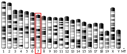
| Location (UCSC) | Chr 11: 85.65 – 85.66 Mb | Chr 7: 90.45 – 90.46 Mb | |||||||||||||||||||||||
| PubMed search | [3] | [4] | |||||||||||||||||||||||
| Wikidata | |||||||||||||||||||||||||
| |||||||||||||||||||||||||

A nonsense mutation in the TMEM126A gene has been shown to be related to optic atrophy. TMEM126A shows higher levels of expression in the parathyroid gland[7] as well as in the peripheral blood cells of Huntington's disease patients, indicating that expression of this protein has some relation to blood regulation.
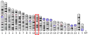
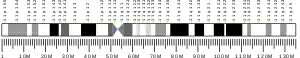
TMEM126A has two isoforms and is found on the long arm of Chromosome 11 in region 1, band 4, sub-band 1.[8] It is produced by the TMEM126 gene, which codes for a mRNA 726 base pairs long.[9] which translates into a protein 195 amino acids long.[10] In addition, this gene is expressed 1.8 times the average of a normal gene and has expression with the prostate, uterus, kidney, placenta, heart, brain, and a large number of other tissues.[11]
Gene
Locus
TMEM126 is located on the long arm of chromosome 11 in humans. It is found on the first sub-band of the fourth band within the first region.[9] This can be written as 11q14.1.
Aliases
TMEM126A is also known as OPA7 and DKFZp586c1924.
Homology/evolutionary history
TMEM126A has been highly conserved among mammals, never dropping below 60% sequence identity when aligned with the same protein sequence from other species. It has been less well conserved outside of mammals, although it is evident in both fish, birds, and some invertebrates.[12]
Orthologs
| Genus and species | Common name | Date of divergence | Accession number | Sequence length | Sequence identity | Sequence similarity | Notes | |
|---|---|---|---|---|---|---|---|---|
| 1 | Gorilla gorilla | Gorilla | 8.8 mya | XP_004051968 | 195 | 98% | 98% | Predicted |
| 2 | Pongo abelii | Sumatran orangutan | 15.7 mya | NP_001127371.1 | 196 | 97% | 98% | Three transmembrane regions |
| 3 | Macaca mulatta | Rhesus macaque | 29.0 mya | NP_001248065 | 195 | 92% | 95% | |
| 4 | Bos Taurus | Cow | 94.2 mya | NP_001032697 | 197 | 81% | 87% | Three transmembrane regions |
| 5 | Canis lupus familiaris | Dog | 94.2 mya | XP_850546 | 197 | 80% | 87% | Predicted |
| 6 | Felis catus | Cat | 94.2 mya | XP_003992716 | 197 | 78% | 87% | Predicted |
| 7 | Sus scrofa | Wild boar | 94.2 mya | NP_001230521 | 196 | 76% | 86% | |
| 8 | Mus musculus | Mouse | 92.3 mya | NP_079736 | 196 | 71% | 84% | Three transmembrane regions |
| 9 | Rattus norvegicus | Brown rat | 92.3 mya | NP_001011557 | 196 | 70% | 81% | Three transmembrane regions |
| 10 | Ornithorhynchus anatinus | Platypus | 167.4 mya | XP_001514960 | 126 | 69% | 82% | |
| 11 | Sarcophilus harrisii | Tasmanian Devil | 162.6 mya | XP_003764651 | 208 | 63% | 80% | Predicted |
| 12 | Salmo salar | Atlantic salmon | 400.1 mya | NP_001134999 | 206 | 47% | 63% | |
| 13 | Gallus gallus | Chicken | 296.0 mya | XP_003640643 | 209 | 46% | 70% | Low quality. Predicted. |
| 14 | Danio rerio | Zebra fish | 400.1 mya | NP_957092 | 201 | 44% | 67% | |
| 15 | Xenopus laevis | African clawed frog | 371.2 mya | NP_001079826 | 203 | 52% | 69% | |
| 16 | Cavia porcellus | Guinea pig | 92.3 mya | XP_003468633 | 195 | 74% | 83% | |
| 17 | Ciona intestinalis | Vase tunicate | 722.5 mya | XP_002124594 | 230 | 28% | 46% |
mRNA
Promoter Analysis
Promoter sequence:
TTCACCCAACACTGCTTCCAAATAAGCAGTACTCTGGAGAACACGAGAAATCCTCAGAAAAATAAGCTGCAGCTCTGAGG
TGCTGATTATGGTAGGGCAATCAATACAGATCAAAACATGGCACAGGGAGCTTAAGTTCCTAGGGAGAGTAGAAAATCGA
TAGAGCCAAGAAATAGCTCACCTTTGACTTATTTTTAACCTGAGTAGCTATCATATGCCAAGAGCTGTGCAGTTTTCATT
TACCCCATGCCAAGAACGTAAGTAGGCTCTACTGACCAGGAAGTTAAGTAATATGCCCGAGGTACGTTTTCAATGGAAGA
GGCTGACTGAGGGTCACCCAACTTATATCTCGAAATTTCACAATTTCTACAAGTTCTGTCCTGGGAGGCAAGAGTAGGTG
AAACGAGCACACTCTACGCCAGGCAAACAAACCTCAACGCTTAGCCTCCCGGCACCTCCTAGGGCCGGAAGCTTCTCAGC
CCAAAGCCGCTGCTGGCTGCAACCTCCGTCCCGCAGTCCAATTAGCAGCCGCGACCCGGCGCCCGCCCACGCCGCGTCAC
GAGTCAGCCAAAGATGGCTGCGCCCAGGTAATTTGAGCAAAGGCCACAGTGAACTCCGGCGTGGCTGAGGAAGGAGGAGG
CACCCACAGGCTGCTGGGAGGAGAGCATAAGGTACTGGTATTCCGGGGGAGGGGGTGAAGTAAATGTCCCGGTGTCAGGA
GAAGCACGACGCGG[15]
| Species | Max Identity |
|---|---|
| Gorilla gorilla | 97% |
| Pan paniscus | 96% |
| Pan troglodytes | 95% |
| Papio anubis | 92% |
| Pango abelii | 92% |
| Macaca mullata | 90% |
There was only one promoter sequence found by using ElDorado on Genomatix. It is 734 base pairs long and found far upstream of the coding region of TMEM126A. The promoter sequence experiences extremely high levels of expression among primates but could not be found in any other species.
mRNA Folding
The minimum free energy formation has a number of hairpin loops and bulges. Another formation exists with a much larger bulge but which maintains the same hairpin loops.
TMEM126A has a number of likely locations for the formation of hairpin loops, four of which are extremely likely.
Alternative Splicing
In some circumstances, the second exon of the TMEM126A mRNA can be spliced out. This exon contains the main starting codon for the gene. The loss of this region would delay the start of translation until the next methionine, which occurs later in exon 3. Ultimately, this causes a loss of a great deal of genetic information from exons 2 and three.
Protein
Isoforms
The protein has three isoforms (a,b and c). The first isoform is the main version and the longest while the other two are both shorter.
Secondary structure
TMEM126A has a mixture of alpha helices, beta strands and coils that make up its secondary structure. There are two regions of high alpha-helix density which correspond to the transmembrane regions of the protein.
Domains and motifs
TMEM126A contains four exons, two transmembrane regions, and three stem-loop capable regions.[16][17]
Post-translational modifications
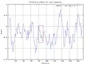
A peptide interaction can be found at amino acid 82 and continues to amino acid 90. This regions has good solubility, thanks to the presence of both cysteine and valine, is not conserved in rabbits, which allows for antibody production, and demonstrates high hydrophobicity.
In addition, there are two glycosylation sites at amino acids 13 and 60.[18] as well as one phosphorylation site at amino acid 40[19]
Expression
TMEM126A is expressed ubiquitously throughout the human body at 1.8 times the normal expression level for human genes. It experiences especially high expression in the parathyroid gland.[7] In addition, TMEM126A experiences higher levels of expression in peripheral blood cells in patients diagnosed with Huntington's disease as well as individuals who experience ocular dominance.
Interacting proteins
TMEM126A interacts with a number of proteins. MYC and MAX form a complex and promote transcription.[20] There is interaction with ATP synthase and the optic atrophy protein.[21] These interactions relate to the proteins function in the mitochondria as well as its medical applications.
Clinical significance
A nonsense mutation in the TMEM126A gene has been shown to be related to optic atrophy.[22] This mutation occurs on the second exon of the protein. The mutation results in decreased expression of TMEM126A. It has been demonstrated that a mutated TMEM126A gene can be distinguished from a normal gene through the use of antibodies which recognize the differences between the two. Experiments have shown that it associates with CD137L in myeloid cells.[23]
Model organisms
Model organisms have been used in the study of TMEM126A function. A conditional knockout mouse line called Tmem126atm1a(EUCOMM)Wtsi was generated at the Wellcome Trust Sanger Institute.[24] Male and female animals underwent a standardized phenotypic screen[25] to determine the effects of deletion.[26][27][28][29] Additional screens performed: - In-depth immunological phenotyping[30]
| Characteristic | Phenotype |
|---|---|
| All data available at.[25][31] | |
| Insulin | Normal |
| Homozygous viability at P14 | Normal |
| Homozygous Fertility | Normal |
| Body weight | Normal |
| Neurological assessment | Normal |
| Grip strength | Normal |
| Dysmorphology | Normal |
| Indirect calorimetry | Normal |
| Glucose tolerance test | Normal |
| Auditory brainstem response | Normal |
| DEXA | Normal |
| Radiography | Normal |
| Eye morphology | Normal |
| Clinical chemistry | Normal |
| Haematology 16 Weeks | Normal |
| Peripheral blood leukocytes 16 Weeks | Normal |
| Heart weight | Normal |
| Salmonella infection | Normal |
| Spleen Immunophenotyping | Normal |
| Mesenteric Lymph Node Immunophenotyping | Normal |
| Epidermal Immune Composition | Abnormal |
| Influenza Challenge | Normal |
References
- GRCh38: Ensembl release 89: ENSG00000171202 - Ensembl, May 2017
- GRCm38: Ensembl release 89: ENSMUSG00000030615 - Ensembl, May 2017
- "Human PubMed Reference:". National Center for Biotechnology Information, U.S. National Library of Medicine.
- "Mouse PubMed Reference:". National Center for Biotechnology Information, U.S. National Library of Medicine.
- "Entrez Gene: transmembrane protein 126A".
- Wiemann S, Weil B, Wellenreuther R, Gassenhuber J, Glassl S, Ansorge W, Böcher M, Blöcker H, Bauersachs S, Blum H, Lauber J, Düsterhöft A, Beyer A, Köhrer K, Strack N, Mewes HW, Ottenwälder B, Obermaier B, Tampe J, Heubner D, Wambutt R, Korn B, Klein M, Poustka A (Mar 2001). "Toward a catalog of human genes and proteins: sequencing and analysis of 500 novel complete protein coding human cDNAs". Genome Research. 11 (3): 422–35. doi:10.1101/gr.GR1547R. PMC 311072. PMID 11230166.
- "EST Profile - Hs.533725".
- "Genecards: TMEM126A".
- "TMEM126A transmembrane protein 126A [Homo sapiens (human)] - Gene - NCBI".
- "transmembrane protein 126A".
- "AceView: TMEM126A".
- "BLAST: Basic Local Alignment Search Tool".
- "TMEM126B transmembrane protein 126B".
- Hanein S, Perrault I, Roche O, Gerber S, Khadom N, Rio M, Boddaert N, Jean-Pierre M, Brahimi N, Serre V, Chretien D, Delphin N, Fares-Taie L, Lachheb S, Rotig A, Meire F, Munnich A, Dufier JL, Kaplan J, Rozet JM (Apr 2009). "TMEM126A, encoding a mitochondrial protein, is mutated in autosomal-recessive nonsyndromic optic atrophy". American Journal of Human Genetics. 84 (4): 493–8. doi:10.1016/j.ajhg.2009.03.003. PMC 2667974. PMID 19327736.
- "ElDorado Introduction".
- "Homo sapiens transmembrane protein 126A (TMEM126A), RefSeqGene on chromosome 11". 2017-10-05. Cite journal requires
|journal=(help) - "transmembrane protein 126A isoform 1 [Homo sapiens]".
- "NetNGlyc 1.0 Server".
- "NetPhos 3.1 Server".
- "2 items (Homo sapiens) - STRING database".
- "STRING: Functional protein association networks".
- "Genecards: transmembrane protein 126A isoform 2".
- Bae JS, Choi JK, Moon JH, Kim EC, Croft M, Lee HW (Dec 2012). "Novel transmembrane protein 126A (TMEM126A) couples with CD137L reverse signals in myeloid cells". Cellular Signalling. 24 (12): 2227–36. doi:10.1016/j.cellsig.2012.07.021. PMC 3466360. PMID 22885069.
- Gerdin AK (2010). "The Sanger Mouse Genetics Programme: high throughput characterisation of knockout mice". Acta Ophthalmologica. 88: 925–7. doi:10.1111/j.1755-3768.2010.4142.x. S2CID 85911512.
- "International Mouse Phenotyping Consortium".
- Skarnes WC, Rosen B, West AP, Koutsourakis M, Bushell W, Iyer V, Mujica AO, Thomas M, Harrow J, Cox T, Jackson D, Severin J, Biggs P, Fu J, Nefedov M, de Jong PJ, Stewart AF, Bradley A (Jun 2011). "A conditional knockout resource for the genome-wide study of mouse gene function". Nature. 474 (7351): 337–42. doi:10.1038/nature10163. PMC 3572410. PMID 21677750.
- Dolgin E (Jun 2011). "Mouse library set to be knockout". Nature. 474 (7351): 262–3. doi:10.1038/474262a. PMID 21677718.
- Collins FS, Rossant J, Wurst W (Jan 2007). "A mouse for all reasons". Cell. 128 (1): 9–13. doi:10.1016/j.cell.2006.12.018. PMID 17218247. S2CID 18872015.
- White JK, Gerdin AK, Karp NA, Ryder E, Buljan M, Bussell JN, Salisbury J, Clare S, Ingham NJ, Podrini C, Houghton R, Estabel J, Bottomley JR, Melvin DG, Sunter D, Adams NC, Tannahill D, Logan DW, Macarthur DG, Flint J, Mahajan VB, Tsang SH, Smyth I, Watt FM, Skarnes WC, Dougan G, Adams DJ, Ramirez-Solis R, Bradley A, Steel KP (Jul 2013). "Genome-wide generation and systematic phenotyping of knockout mice reveals new roles for many genes". Cell. 154 (2): 452–64. doi:10.1016/j.cell.2013.06.022. PMC 3717207. PMID 23870131.
- "Infection and Immunity Immunophenotyping (3i) Consortium".
- "Infection and Immunity Immunophenotyping (3i) Consortium".