Valine
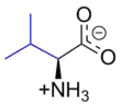
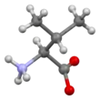
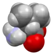
Valine (symbol Val or V)[3] is an α-amino acid that is used in the biosynthesis of proteins. It contains an α-amino group (which is in the protonated −NH3+ form under biological conditions), an α-carboxylic acid group (which is in the deprotonated −COO− form under biological conditions), and a side chain isopropyl group, making it a non-polar aliphatic amino acid. It is essential in humans, meaning the body cannot synthesize it: it must be obtained from the diet. Human dietary sources are foods that contain protein, such as meats, dairy products, soy products, beans and legumes. It is encoded by all codons starting with GU (GUU, GUC, GUA, and GUG).
| |||
| |||
| Names | |||
|---|---|---|---|
| IUPAC name
Valine | |||
| Other names
2-Amino-3-methylbutanoic acid | |||
| Identifiers | |||
3D model (JSmol) |
|||
| ChEBI | |||
| ChEMBL | |||
| ChemSpider | |||
| DrugBank | |||
| ECHA InfoCard | 100.000.703 | ||
| EC Number |
| ||
| KEGG | |||
PubChem CID |
|||
| UNII |
| ||
CompTox Dashboard (EPA) |
|||
| |||
| |||
| Properties[1] | |||
| C5H11NO2 | |||
| Molar mass | 117.148 g·mol−1 | ||
| Density | 1.316 g/cm3 | ||
| Melting point | 298 °C (568 °F; 571 K) (decomposition) | ||
| soluble | |||
| Acidity (pKa) | 2.32 (carboxyl), 9.62 (amino)[2] | ||
| -74.3·10−6 cm3/mol | |||
| Supplementary data page | |||
| Refractive index (n), Dielectric constant (εr), etc. | |||
Thermodynamic data |
Phase behaviour solid–liquid–gas | ||
| UV, IR, NMR, MS | |||
Except where otherwise noted, data are given for materials in their standard state (at 25 °C [77 °F], 100 kPa). | |||
| Infobox references | |||

Like leucine and isoleucine, valine is a branched-chain amino acid. In sickle-cell disease, a single glutamic acid in β-globin is replaced with valine. Because valine is hydrophobic, whereas glutamic acid is hydrophilic, this change makes the hemoglobin prone to abnormal aggregation.
History and etymology
Valine was first isolated from casein in 1901 by Hermann Emil Fischer.[4] The name valine comes from valeric acid, which in turn is named after the plant valerian due to the presence of the acid in the roots of the plant.[5][6]
Nomenclature
According to IUPAC, carbon atoms forming valine are numbered sequentially starting from 1 denoting the carboxyl carbon, whereas 4 and 4' denote the two terminal methyl carbons.[7]
Metabolism
Source and biosynthesis
Valine, like other branched-chain amino acids, is synthesized by plants, but not by animals.[8] It is therefore an essential amino acid in animals, and needs to be present in the diet. Adult humans require about 4 mg/kg body weight daily.[9] It is synthesized in plants and bacteria via several steps starting from pyruvic acid. The initial part of the pathway also leads to leucine. The intermediate α-ketoisovalerate undergoes reductive amination with glutamate. Enzymes involved in this biosynthesis include:[10]
- Acetolactate synthase (also known as acetohydroxy acid synthase)
- Acetohydroxy acid isomeroreductase
- Dihydroxyacid dehydratase
- Valine aminotransferase
Degradation
Like other branched-chain amino acids, the catabolism of valine starts with the removal of the amino group by transamination, giving alpha-ketoisovalerate, an alpha-keto acid, which is converted to isobutyryl-CoA through oxidative decarboxylation by the branched-chain α-ketoacid dehydrogenase complex.[11] This is further oxidised and rearranged to succinyl-CoA, which can enter the citric acid cycle.
Synthesis
Racemic valine can be synthesized by bromination of isovaleric acid followed by amination of the α-bromo derivative[12]
Medical significance
Insulin resistance
Valine, like other branched-chain amino acids, is associated with insulin resistance: higher levels of valine are observed in the blood of diabetic mice, rats, and humans.[13] Mice fed a valine deprivation diet for one day have improved insulin sensitivity, and feeding of a valine deprivation diet for one week significantly decreases blood glucose levels.[14] In diet-induced obese and insulin resistant mice, a diet with decreased levels of valine and the other branched-chain amino acids results in reduced adiposity and improved insulin sensitivity.[15] The valine catabolite 3-hydroxyisobutyrate promotes skeletal muscle insulin resistance in mice by stimulating fatty acid uptake into muscle and lipid accumulation.[16] In humans, a protein restricted diet lowers blood levels of valine and decreases fasting blood glucose levels.[17]
Hematopoietic stem cells
Dietary valine is essential for hematopoietic stem cell (HSC) self-renewal, as demonstrated by experiments in mice.[18] Dietary valine restriction selectively depletes long-term repopulating HSC in mouse bone marrow. Successful stem cell transplantation was achieved in mice without irradiation after 3 weeks on a valine restricted diet. Long term survival of the transplanted mice was achieved when valine was returned to the diet gradually over a 2-week period to avoid refeeding syndrome.
See also
References
- Weast, Robert C., ed. (1981). CRC Handbook of Chemistry and Physics (62nd ed.). Boca Raton, FL: CRC Press. p. C-569. ISBN 0-8493-0462-8.
- Dawson, R.M.C., et al., Data for Biochemical Research, Oxford, Clarendon Press, 1959.
- "Nomenclature and Symbolism for Amino Acids and Peptides". IUPAC-IUB Joint Commission on Biochemical Nomenclature. 1983. Archived from the original on 9 October 2008. Retrieved 5 March 2018.
- "valine". Encyclopædia Britannica Online. Retrieved 6 December 2015.
- "valine". Merriam-Webster Online Dictionary. Retrieved 6 December 2015.
- "valeric acid". Merriam-Webster Online Dictionary. Retrieved 6 December 2015.
- Jones, J. H., ed. (1985). Amino Acids, Peptides and Proteins. Specialist Periodical Reports. 16. London: Royal Society of Chemistry. p. 389. ISBN 978-0-85186-144-9.
- Basuchaudhuri, Pranab (2016). Nitrogen metabolism in rice. Boca Raton, Florida: CRC Press. p. 159. ISBN 9781498746687. OCLC 945482059.
- Institute of Medicine (2002). "Protein and Amino Acids". Dietary Reference Intakes for Energy, Carbohydrates, Fiber, Fat, Fatty Acids, Cholesterol, Protein, and Amino Acids. Washington, DC: The National Academies Press. pp. 589–768.
- Lehninger, Albert L.; Nelson, David L.; Cox, Michael M. (2000). Principles of Biochemistry (3rd ed.). New York: W. H. Freeman. ISBN 1-57259-153-6..
- Mathews, Christopher K. (2000). Biochemistry. Van Holde, K. E., Ahern, Kevin G. (3rd ed.). San Francisco, Calif.: Benjamin Cummings. p. 776. ISBN 0805330666. OCLC 42290721.
- Marvel, C. S. (1940). "dl-Valine". Organic Syntheses. 20: 106.; Collective Volume, 3, p. 848.
- Lynch, Christopher J.; Adams, Sean H. (1 December 2014). "Branched-chain amino acids in metabolic signalling and insulin resistance". Nature Reviews. Endocrinology. 10 (12): 723–736. doi:10.1038/nrendo.2014.171. ISSN 1759-5037. PMC 4424797. PMID 25287287.
- Xiao, Fei; Yu, Junjie; Guo, Yajie; Deng, Jiali; Li, Kai; Du, Ying; Chen, Shanghai; Zhu, Jianmin; Sheng, Hongguang (1 June 2014). "Effects of individual branched-chain amino acids deprivation on insulin sensitivity and glucose metabolism in mice". Metabolism: Clinical and Experimental. 63 (6): 841–850. doi:10.1016/j.metabol.2014.03.006. ISSN 1532-8600. PMID 24684822.
- Cummings, Nicole E.; Williams, Elizabeth M.; Kasza, Ildiko; Konon, Elizabeth N.; Schaid, Michael D.; Schmidt, Brian A.; Poudel, Chetan; Sherman, Dawn S.; Yu, Deyang (19 December 2017). "Restoration of metabolic health by decreased consumption of branched-chain amino acids". The Journal of Physiology. 596 (4): 623–645. doi:10.1113/JP275075. ISSN 1469-7793. PMC 5813603. PMID 29266268.
- Jang, Cholsoon; Oh, Sungwhan F.; Wada, Shogo; Rowe, Glenn C.; Liu, Laura; Chan, Mun Chun; Rhee, James; Hoshino, Atsushi; Kim, Boa (1 April 2016). "A branched-chain amino acid metabolite drives vascular fatty acid transport and causes insulin resistance". Nature Medicine. 22 (4): 421–426. doi:10.1038/nm.4057. ISSN 1546-170X. PMC 4949205. PMID 26950361.
- Fontana, Luigi; Cummings, Nicole E.; Arriola Apelo, Sebastian I.; Neuman, Joshua C.; Kasza, Ildiko; Schmidt, Brian A.; Cava, Edda; Spelta, Francesco; Tosti, Valeria (21 June 2016). "Decreased Consumption of Branched-Chain Amino Acids Improves Metabolic Health". Cell Reports. 16 (2): 520–30. doi:10.1016/j.celrep.2016.05.092. ISSN 2211-1247. PMC 4947548. PMID 27346343.
- Taya, Yuki; Ota, Yasunori; Wilkinson, Adam C.; Kanazawa, Ayano; Watarai, Hiroshi; Kasai, Masataka; Nakauchi, Hiromitsu; Yamazaki, Satoshi (2 December 2016). "Depleting dietary valine permits nonmyeloablative mouse hematopoietic stem cell transplantation". Science. 354 (6316): 1152–1155. Bibcode:2016Sci...354.1152T. doi:10.1126/science.aag3145. PMID 27934766. S2CID 45815137.
External links
| General topics |  | ||||||||||
|---|---|---|---|---|---|---|---|---|---|---|---|
| By properties |
| ||||||||||
| |||||||||||