Vagal trigone
The cells of the dorsal nucleus of vagus nerve are spindle-shaped, like those of the posterior column of the spinal cord, and the nucleus is usually considered as representing the base of the posterior column. It measures about 2 cm. in length, and in the lower, closed part of the medulla oblongata is situated behind the dorsal motor nucleus of the vagus; whereas in the upper, open part it lies lateral to that nucleus, and corresponds to an eminence, named the vagal trigone (ala cinerea, not to be confused with tuberculum cinereum nor tuber cinereum), in the rhomboid fossa.
| Vagal trigone | |
|---|---|
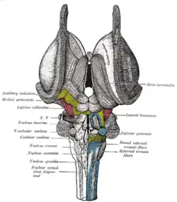 Dissection of brain-stem. Dorsal view. | |
 Human caudal brainstem posterior view (Trigonum nervi vagi is #7) | |
| Details | |
| Identifiers | |
| Latin | trigonum nervi vagi |
| NeuroNames | 634 |
| TA98 | A14.1.05.709 |
| FMA | 78445 |
| Anatomical terms of neuroanatomy | |
The vagal trigone is separated from the area postrema by a narrow strip of thickened ependyma – the funiculus separans.[1]
References
- Mirza M, M Das J (8 July 2019). Neuroanatomy, Area Postrema. StatPearls; Treasure Island (FL): StatPearls Publishing. PMID 31334969.CS1 maint: uses authors parameter (link)
This article incorporates text in the public domain from page 781 of the 20th edition of Gray's Anatomy (1918)
External links
| Authority control |
|---|