Advanced airway management
Advanced airway management is the subset of airway management that involves advanced training, skill, and invasiveness. It encompasses various techniques performed to create an open or patent airway – a clear path between a patient's lungs and the outside world.
| Advanced airway management | |
|---|---|
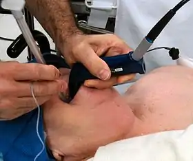 Photograph of an anesthesiologist using the Glidescope video laryngoscope to intubate the trachea of a morbidly obese elderly person with challenging airway anatomy |
This is accomplished by clearing or preventing obstructions of airways. Obstructions can be caused by many things, including the patient's own tongue or other anatomical components of the airway, foreign bodies, excessive amounts of blood and body fluids, or aspiration of food particles.
Unlike basic airway management such as Head tilt/Chin lift or jaw-thrust maneuver, advanced airway management relies on the use of medical equipment and advanced training. Certain invasive airway management techniques can be performed "blind" or with visualization of the glottis. Visualization of the glottis can be accomplished either directly by using a laryngoscope blade or by utilizing newer video technology options.
In roughly increasing order of invasiveness are the use of supraglottic devices such as oropharyngeal (OPA), nasopharyngeal (NPA), and laryngeal mask airways (LMA). Laryngeal mask airways can even be used to deliver general anesthesia. These are followed by infraglottic techniques, such as tracheal intubation and finally surgical techniques.
Advanced airway management is a key component in cardiopulmonary resuscitation, anaesthesia, emergency medicine, and intensive care medicine. The A in the ABC initialism mnemonic for dealing with critically ill patients stands for airway management. Many airways are straightforward to manage. However, some can be challenging. Such difficulties can be predicted to some extent; a recent Cochrane systematic review examines the sensitivity and specificity of the various bedside tests commonly used to predict difficulty in airway management.[1]
Removal of foreign objects
In advanced airway management foreign objects are either removed by suction or with e.g. a Magill forceps under inspection of the airway with a laryngoscope or bronchoscope. If removal is not possible surgical methods should be considered.
Supraglottic techniques
Supraglottic techniques include the use of supraglottic tubes, such as oropharyngeal and nasopharyngeal airways, and supraglottic devises such as laryngeal masks. Common for all supraglottic devises are that they are introduced into the pharynx, ensuring the upper respiratory tract remains open, without passing through the glottis and thereby entering the trachea.
Nasopharyngeal airway
Nasopharyngeal airways is a soft rubber or plastic hollow tube that is passed through the nose into the posterior pharynx. Patients tolerate NPAs more easily than OPAs, so NPAs can be used when the use of an OPA is difficult, such as when the patient's jaw is clenched or the patient is semiconscious and cannot tolerate an OPA.[2] NPAs are generally not recommended if there is suspicion of a fracture to the base of the skull, due to the possibility of the tube entering the cranium.[3] However, the actual risks of this complication occurring compared to the risks of damage from hypoxia if an airway is not used are debatable.[3][4]
Oropharyngeal airway

Oropharyngeal airways are rigid plastic curved devices, which are inserted through the patients mouth. It prevents the patients tongue from covering the epiglottis and thereby obstructing the airway. An oropharyngeal airway should only be used in a deeply unresponsive patient because in a responsive patient they can cause vomiting and aspiration by stimulating the gag reflex.[5]
Supraglottic airway
Supraglottic airways (or extraglottic devices[6]) are a family of devices that are inserted through the mouth to sit on top of the larynx. Supraglottic airways are used in the majority of operations performed under general anaesthesia.[7] Compared to a cuffed tracheal tube (see below), they give less protection against aspiration but are easier to insert and cause less laryngeal trauma.[6]
The best-known example is the laryngeal mask airway. A laryngeal mask airway is an airway placed into the mouth and set over the glottis and inflated.[8] Other variations include devices with oesophageal access ports, so that a separate tube can be inserted from the mouth to the stomach to decompress accumulated gases and drain liquid contents.[6] Some devices can have an endotracheal tube passed through them into the trachea.[6]
Infraglottic techniques
Indications
There are specific indications or guidelines for deciding a more invasive and more secure airway is worth the associated risk:[9]
- respiratory failure
- apnea or the suspension of breathing
- decreased or altered level of consciousness, rapid mental status change, Glasgow Coma Scale score less than 8 (GCS<8).
- major trauma, such as penetrating injury to abdomen or chest
- direct airway injury or facial burns
- high risk of aspiration
Tracheal intubation
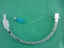
Tracheal intubation, often simply referred to as intubation, is the placement of a flexible plastic or rubber tube into the trachea to maintain an open airway or to serve as a conduit through which to administer certain drugs. It is frequently performed in critically injured, ill or anesthetized patients to facilitate ventilation of the lungs, including mechanical ventilation, and to prevent the possibility of asphyxiation or airway obstruction. The most widely used route is orotracheal, in which an endotracheal tube is passed through the mouth and vocal apparatus into the trachea. In a nasotracheal procedure, an endotracheal tube is passed through the nose and vocal apparatus into the trachea.
Classically tracheal intubation has been performed utilizing laryngoscopic blades to obtain direct visualization of the vocal cords. Even in this category there are multiple different blade styles, shapes and lengths from which to choose. Multiple intubation tools are now available with built-in video technology. A Glidescope utilizes a laryngoscopic blade connected by a cable to a large video screen and requires a slightly different technique than that of a traditional laryngoscope. The McGrath model has a compact design with a smaller screen directly attached to the blade. Studies have shown that video laryngoscopes when compared to classic models resulted in fewer failed intubation attempts, especially in those patients designated as more difficult airways. These devices are quickly finding their way into emergency departments, operating theaters and critical care floors across the world.[10]
Alternatives to standard endotracheal tubes includes laryngeal tube and combitube.
Confirming placement
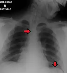
The absolute gold standard for confirming successful placement of an endotracheal tube is direct visualization of the tube passing through the vocal cords. Other methods used as secondary confirmation include carbon dioxide detectors, capnography, oxygen saturation, chest x-ray, or equal chest rise and breath sounds heard on both sides of the chest.[9]
Surgical methods
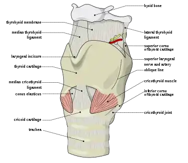
Surgical methods for airway management rely on making a surgical incision is made below the glottis in order to achieve direct access to the lower respiratory tract, bypassing the upper respiratory tract. Surgical airway management is often performed as a last resort in cases where orotracheal and nasotracheal intubation are impossible or contraindicated. Surgical airway management is also used when a person will need a mechanical ventilator for a longer period. Surgical methods for airway management include cricothyrotomy and tracheostomy.
A cricothyrotomy is an incision made through the skin and cricothyroid membrane to establish a patent airway during certain life-threatening situations, such as airway obstruction by a foreign body, angioedema, or massive facial trauma.[11] A cricothyrotomy is nearly always performed as a last resort in cases where orotracheal and nasotracheal intubation are impossible or contraindicated. Cricothyrotomy is easier and quicker to perform than tracheotomy, does not require manipulation of the cervical spine, and is associated with fewer complications.[12]
A tracheotomy is a surgically created opening from the skin of the neck down to the trachea.[13] A tracheotomy may be considered where a person will need to be on a mechanical ventilator for a longer period.[13] The advantages of a tracheotomy include less risk of infection and damage to the trachea such as tracheal stenosis.[13]
Pediatric considerations
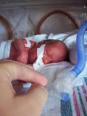
Children are not just small adults. They are unique in far more ways than simply being smaller in size. There are many basic differences in anatomy compared to adults that can affect airway management. For example, children's heads are proportionally larger in relation to their overall body size. This can cause alignment issues that have the potential to make it substantially more difficult to obtain good visualization of the appropriate airway landmarks. The differences in a child's anatomy can also affect equipment choices, such as choosing a straight laryngoscope blade instead of a curved one to achieve better control of a more elastic airway. Making the right equipment choices is so important that a color-coded tape measure (known as Broselow tape) was created to help facilitate rapid and accurate decisions in pediatric emergency situations. Birth complications, congenital syndromes (such as Down syndrome) and even recent illness or nasal congestion can affect how airway management is approached in a child.
When ventilation, various airway options and even intubation are unsuccessful, this is a terrifying situation known as "cannot ventilate, cannot intubate". Typically this is when a cricothyrotomy would be attempted as mentioned above. However, this tricky procedure is even more difficult in kids due to their extra flexible airways. The chance of accidentally puncturing all the way through the trachea to the esophagus increases substantially. The risk is considered so high that the procedure is contraindicated in children under the age of 5–6 years old.[14]
See also
References
- Roth, Dominik; et al. (2018). "Airway physical examination tests for detection of difficult airway management in apparently normal adult patients". Cochrane Database of Systematic Reviews. 2018 (5): CD008874. doi:10.1002/14651858.CD008874.pub2. PMC 6404686. PMID 29761867.
- Roberts K, Whalley H, Bleetman A (2005). "The nasopharyngeal airway: dispelling myths and establishing the facts". Emerg Med J. 22 (6): 394–6. doi:10.1136/emj.2004.021402. PMC 1726817. PMID 15911941.
- Ellis, D. Y. (2006). "Intracranial placement of nasopharyngeal airways: Is it all that rare?". Emergency Medicine Journal. 23 (8): 661. doi:10.1136/emj.2006.036541. PMC 2564185. PMID 16858116.
- Roberts, K.; Whalley, H.; Bleetman, A. (2005). "The nasopharyngeal airway: Dispelling myths and establishing the facts". Emergency Medicine Journal. 22 (6): 394–396. doi:10.1136/emj.2004.021402. PMC 1726817. PMID 15911941.
- "Guedel airway". AnaesthesiaUK. 14 May 2010. Archived from the original on 24 January 2013. Retrieved 23 May 2013.
- Hernandez, MR; Klock, A; Ovassapian, A (2011). "Evolution of the Extraglottic Airway: A Review of Its History, Applications, and Practical Tips for Success". Anesthesia and Analgesia. 114 (2): 349–68. doi:10.1213/ANE.0b013e31823b6748. PMID 22178627.
- Cook, T; Howes, B. (2010). "Supraglottic airway devices: recent advances". Continuing Education in Anaesthesia, Critical Care and Pain. 11 (2): 56. doi:10.1093/bjaceaccp/mkq058.
- Davies PR, Tighe SQ, Greenslade GL, Evans GH (1990). "Laryngeal mask airway and tracheal tube insertion by unskilled personnel". The Lancet. 336 (8721): 977–979. doi:10.1016/0140-6736(90)92429-L. PMID 1978159. Retrieved 25 July 2010.
- Avva U, Bhimji SS. Airway, Management. [Updated 2017 Dec 15]. In: StatPearls [Internet]. Treasure Island (FL): StatPearls Publishing; 2018 Jan-. Available from: https://www.ncbi.nlm.nih.gov/books/NBK470403/
- Lewis SR, Butler AR, Parker J, Cook TM, Smith AF. Videolaryngoscopy versus direct laryngoscopy for adult patients requiring tracheal intubation. Cochrane Database of Systematic Reviews 2016, Issue 11. Art. No: CD011136 DOI: 10.1002/14651858.CD011136.pub2.
- Mohan, R; Iyer, R; Thaller, S (2009). "Airway management in patients with facial trauma". Journal of Craniofacial Surgery. 20 (1): 21–3. doi:10.1097/SCS.0b013e318190327a. PMID 19164982.
- Katos, MG; Goldenberg, D (2007). "Emergency cricothyrotomy". Operative Techniques in Otolaryngology. 18 (2): 110–4. doi:10.1016/j.otot.2007.05.002.
- Andriolo, Brenda N. G.; Andriolo, Régis B.; Saconato, Humberto; Atallah, Álvaro N.; Valente, Orsine (2015-01-12). "Early versus late tracheostomy for critically ill patients". The Cochrane Database of Systematic Reviews. 1: CD007271. doi:10.1002/14651858.CD007271.pub3. ISSN 1469-493X. PMC 6517297. PMID 25581416.
- Harless J, Ramalah R, Bhananker SM. Pediatric airway management. Int J Crit Illn Inj Sci 2014; 4:65–70.