Cervical cancer staging
Cervical cancer staging is the assessment of cervical cancer to decide how far the disease has progressed. Cancer staging generally runs from stage 0, which is pre-cancerous or non-invasive, to stage IV, in which the cancer has spread throughout a significant part of the body.[1]
Cervical cancer is staged by the International Federation of Gynecology and Obstetrics (FIGO) staging system.[2] Prior to the 2018 update, FIGO staging of cervical cancer allowed only the following diagnostic tests to be used in determining the stage: palpation (feeling with the fingers), inspection, colposcopy, endocervical curettage, hysteroscopy, cystoscopy, proctoscopy, intravenous urography, and X-ray examination of the lungs and skeleton, and cervical conization. But with the 2018 update of FIGO staging of cervical cancer, imaging was allowed to assess primary tumor size and/or lymph node status of patients clinical stages I through III disease.[3]
Stages
 Stage 1A cervical cancer
 Stage 1B cervical cancer
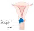 Stage 2A cervical cancer
 Stage 2B cervical cancer
 Stage 3B cervical cancer
 Stage 4A cervical cancer
 Stage 4B cervical cancer
Stage 0
- The carcinoma is confined to the surface layer (cells lining) of the cervix. Also called carcinoma in situ (CIS).
Stage I
- The carcinoma has grown into the cervix, but has not spread beyond it (extension to the corpus would be disregarded). Stage One is subdivided as follows:
- IA: Invasive carcinoma which can be diagnosed only by microscopy, with deepest invasion <5 mm
- IA1: Measured stromal invasion <3.0 mm
- IA2: Measured stromal invasion ≥3.0 mm and <5 mm
- IB: Invasive carcinoma with measured deepest invasion ≥5 mm, limited to the cervix
- IB1: Invasive carcinoma ≥5 mm depth of invasion and <2 cm in greatest dimension
- IB2: Invasive carcinoma ≥2 cm and <4 cm in greatest dimension
- IB3: Invasive carcinoma ≥4.0 cm in greatest dimension
- IA: Invasive carcinoma which can be diagnosed only by microscopy, with deepest invasion <5 mm
Stage II
- Cervical carcinoma invades beyond the uterus, but not to the pelvic wall or to the lower third of the vagina
- IIA: Without parametrial invasion
- IIA1: Tumor <4.0 cm in greatest dimension
- IIA2: Tumor ≥4.0 cm in greatest dimension
- IIB: With parametrial invasion
- IIA: Without parametrial invasion
Stage III
- The carcinoma involves the lower third of the vagina and/or extends to the pelvic wall and/or causes hydronephrosis or non‐functioning kidney and/or involves pelvic and/or paraaortic lymph nodes.
- IIIA: Carcinoma involves the lower third of the vagina, with no extension to the pelvic wall.
- IIIB: Extension to the pelvic wall and/or hydronephrosis or non‐functioning kidney.
- IIIC: Involvement of pelvic and/or paraaortic lymph nodes, irrespective of tumor size and extent
- IIIC1: Pelvic lymph node metastasis only
- IIIC2: Para‐aortic lymph node metastasis
Stage IV
- The carcinoma has extended beyond the true pelvis or has involved (biopsy proven) the mucosa of the bladder or rectum. A bullous edema, as such, does not permit a case to be allotted to Stage IV
- IVA: Spread of the growth to adjacent organs
- IVB: Spread to distant organs[3]
References
- "Staging". National Cancer Institute. Retrieved 2018-11-07.
- "Cervical Cancer Stages". www.cancer.org. Retrieved 2021-01-02.
- Bhatla, Neerja; Berek, Jonathan S.; Fredes, Mauricio Cuello; Denny, Lynette A.; Grenman, Seija; Karunaratne, Kanishka; Kehoe, Sean T.; Konishi, Ikuo; Olawaiye, Alexander B.; Prat, Jaime; Sankaranarayanan, Rengaswamy (2019). "Revised FIGO staging for carcinoma of the cervix uteri". International Journal of Gynecology & Obstetrics. 145 (1): 129–135. doi:10.1002/ijgo.12749. ISSN 1879-3479.