Embryonic disc
The embryonic disc (or embryonic disk) forms the floor of the amniotic cavity. It is composed of a layer of prismatic cells – the embryonic ectoderm, derived from the inner cell mass and lying in apposition with the endoderm.
| Embryonic disc | |
|---|---|
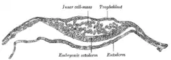 Section through embryonic disc of a parti-coloured bat. | |
 | |
| Details | |
| Carnegie stage | 4 |
| Precursor | Ectoderm |
| Identifiers | |
| Latin | discus embryonicus |
| TE | E2.0.1.2.0.0.14 |
| Anatomical terminology | |
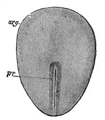
In humans, it is the stage of development that occurs after implantation and prior to the embryonic folding (e.g. seen between about day 14 to day 21 post fertilization). It is derived from the epiblast layer, which lies between the hypoblast layer and the amnion.[1] The epiblast layer is derived from the inner cell mass.[2] Through the process of gastrulation, the bilaminar embryonic disc becomes trilaminar. After this, the notochord forms. Through the process of neurulation, the notochord induces the formation of the neural tube in the embryonic disc.
References
This article incorporates text in the public domain from page 47 of the 20th edition of Gray's Anatomy (1918)
- Tan, Wen-Hann; Gilmore, Edward C.; Baris, Hagit N. (2013-01-01), Rimoin, David; Pyeritz, Reed; Korf, Bruce (eds.), "Chapter 15 - Human Developmental Genetics", Emery and Rimoin's Principles and Practice of Medical Genetics, Oxford: Academic Press, pp. 1–63, doi:10.1016/b978-0-12-383834-6.00018-5, ISBN 978-0-12-383834-6, retrieved 2020-11-13
- Neidhart, Michel (2016-01-01), Neidhart, Michel (ed.), "Chapter 13 - DNA Methylation and Development", DNA Methylation and Complex Human Disease, Oxford: Academic Press, pp. 229–240, doi:10.1016/b978-0-12-420194-1.00013-0, ISBN 978-0-12-420194-1, retrieved 2020-11-13