Infrahyoid muscles
The infrahyoid muscles, or strap muscles, are a group of four pairs of muscles in the anterior (frontal) part of the neck.[1] The four infrahyoid muscles are the sternohyoid, sternothyroid, thyrohyoid and omohyoid muscles.[1]
| Infrahyoid muscles | |
|---|---|
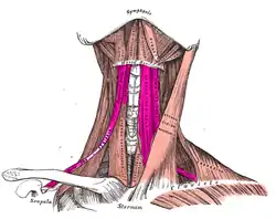 Muscles of the neck seen from the front. The infrahyoid muscles are coloured in violet. | |
| Details | |
| Nerve | Ansa cervicalis |
| Actions | Depress the hyoid bone |
| Identifiers | |
| Latin | Musculi infrahyoidei |
| TA98 | A04.2.04.001 |
| TA2 | 2167 |
| FMA | 71298 |
| Anatomical terms of muscle | |
Excluding the sternothyroid, the infrahyoid muscles either originate from or insert on to the hyoid bone.[2]
The term infrahyoid refers to the region below the hyoid bone, while the term strap muscles refers to the long and flat muscle shapes which resembles a strap. The stylopharyngeus muscle is considered by many to be one of the strap muscles, but is not an infrahyoid muscle.
Individual muscles
The origin, insertion and innervation of the individual muscles:[3]
| Muscle | Origin | Insertion | Innervation |
| Sternohyoid | Posterior surface of manubrium sterni, adjoining parts of clavicle and the posterior sternoclavicular ligament | Medial part of lower border of hyoid bone | Ansa cervicalis |
| Sternothyroid | Posterior surface of manubrium sterni and adjoining part of first costal cartilage | Oblique line of thyroid cartilage | Ansa cervicalis |
| Thyrohyoid | Oblique line of the thyroid cartilage | Lower border of the body and the greater cornu of the hyoid bone | Cervical spinal nerve 1 via the hypoglossal nerve |
| Omohyoid (superior belly) | Intermediate tendon | Hyoid bone | Superior root of ansa cervicalis (C1) |
| Omohyoid (inferior belly) | Superior border of scapula | Intermediate tendon | Ansa cervicalis (C1-C3) |
Nerve supply
All of the infrahyoid muscles are innervated by the ansa cervicalis from the cervical plexus (C1-C3)[4][5] except the thyrohyoid muscle, which is innervated by fibers only from the first cervical spinal nerve travelling with the hypoglossal nerve.[1]
Function
The infrahyoid muscles function to elevate and depress the hyoid bone and larynx during swallowing and speech.[6] This moves the larynx as one unit.[7]
See also
References
- "Localization of Motoneurons in the Spinal Cord". The Spinal Cord: 94–114. 1 January 2009. doi:10.1016/B978-0-12-374247-6.50011-0.
- KenHub. "Infrahyoid muscles". Retrieved 24 May 2019.
- Ellis, Harold; Susan Standring; Gray, Henry David (2005). Gray's anatomy: the anatomical basis of clinical practice. St. Louis, Mo: Elsevier Churchill Livingstone. pp. 538–539. ISBN 0-443-07168-3.
- Cesmebasi, Alper (1 January 2015), Tubbs, R. Shane; Rizk, Elias; Shoja, Mohammadali M.; Loukas, Marios (eds.), "Chapter 31 - Anatomy of the Cervical Plexus and Its Branches", Nerves and Nerve Injuries, San Diego: Academic Press, pp. 441–449, doi:10.1016/b978-0-12-410390-0.00032-9, ISBN 978-0-12-410390-0, retrieved 10 November 2020
- Kayalioglu, Gulgun (1 January 2009), Watson, Charles; Paxinos, George; Kayalioglu, Gulgun (eds.), "Chapter 4 - The Spinal Nerves", The Spinal Cord, San Diego: Academic Press, pp. 37–56, doi:10.1016/b978-0-12-374247-6.50008-0, ISBN 978-0-12-374247-6, retrieved 10 November 2020
- Merea, Valeria Silva; Pitman, Michael J. (1 January 2019), Chhetri, Dinesh K.; Dewan, Karuna (eds.), "Chapter 5 - Anatomy and Physiology of the Upper Esophageal Sphincter", Dysphagia Evaluation and Management in Otolaryngology, Elsevier, pp. 29–34, doi:10.1016/b978-0-323-56930-9.00005-x, ISBN 978-0-323-56930-9, retrieved 10 November 2020
- Feinstein, Aaron J.; Dewan, Karuna (1 January 2019), Chhetri, Dinesh K.; Dewan, Karuna (eds.), "Chapter 4 - Larynx", Dysphagia Evaluation and Management in Otolaryngology, Elsevier, pp. 23–28, doi:10.1016/b978-0-323-56930-9.00004-8, ISBN 978-0-323-56930-9, retrieved 10 November 2020
| Authority control |
|---|