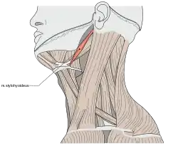
Stylohyoid muscle
The stylohyoid muscle is a slender muscle, lying anterior and superior of the posterior belly of the digastric muscle.[1] It is one of the suprahyoid muscles.[2] It shares this muscle's innervation by the facial nerve,[3] and functions to draw the hyoid bone backwards and elevate the tongue. Its origin is the styloid process of the temporal bone. It inserts on the body of the hyoid.[1]
| Stylohyoid | |
|---|---|
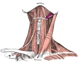
 The stylohyoid among the triangles of the neck. | |
 Muscles of the neck. Anterior view. Stylohyoid muscle in purple | |
| Details | |
| Origin | styloid process (temporal) |
| Insertion | Greater cornu of hyoid bone |
| Nerve | facial nerve (CN VII) |
| Actions | Elevate the hyoid during swallowing |
| Identifiers | |
| Latin | musculus stylohyoideus |
| TA98 | A04.2.03.005 |
| TA2 | 2164 |
| FMA | 9625 |
| Anatomical terms of muscle | |
Structure
The stylohyoid muscle originates from the posterior and lateral surface of the styloid process of the temporal bone, near the base. Passing inferior and anterior, it inserts into the body of the hyoid bone, at its junction with the greater cornu, and just superior to the omohyoid muscle. It belongs to the group of suprahyoid muscles.[2]
It is perforated, near its insertion, by the intermediate tendon of the digastric muscle.
The stylohyoid muscle has vascular supply from the lingual artery, a branch of the external carotid artery.[4]
Nerve supply
A branch of the facial nerve (CN VII) innervates the stylohyoid muscle.[3][5]
Variation
It may be absent or doubled, lie beneath the carotid artery, or be inserted into the omohyoid, or mylohyoid muscles.
Function
The stylohyoid muscle elevates and retracts hyoid bone. It initiates a swallowing action by pulling the hyoid bone in a posterior and superior direction.
Additional images
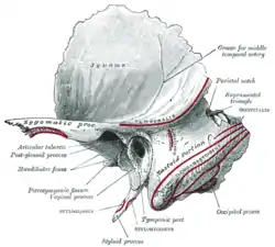 Left temporal bone. Outer surface.
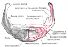 Hyoid bone. Anterior surface. Enlarged.
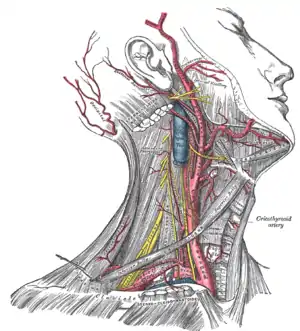 Superficial dissection of the right side of the neck, showing the carotid and subclavian arteries.
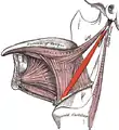 Extrinsic muscles of the tongue. Left side.
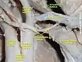 Stylohyoid muscle
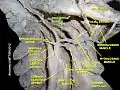 Stylohyoid muscle
See also
References
This article incorporates text in the public domain from page 392 of the 20th edition of Gray's Anatomy (1918)
- Rea, Paul (2016-01-01), Rea, Paul (ed.), "Chapter 2 - Head", Essential Clinically Applied Anatomy of the Peripheral Nervous System in the Head and Neck, Academic Press, pp. 21–130, doi:10.1016/b978-0-12-803633-4.00002-8, ISBN 978-0-12-803633-4, retrieved 2020-11-11
- Chokroverty, Sudhansu (2009-01-01), Chokroverty, Sudhansu (ed.), "Chapter 7 - Physiologic Changes in Sleep", Sleep Disorders Medicine (Third Edition), Philadelphia: W.B. Saunders, pp. 80–104, doi:10.1016/b978-0-7506-7584-0.00007-0, ISBN 978-0-7506-7584-0, retrieved 2020-11-11
- Felten, David L.; O'Banion, M. Kerry; Maida, Mary Summo (2016-01-01), Felten, David L.; O'Banion, M. Kerry; Maida, Mary Summo (eds.), "11 - Brain Stem and Cerebellum", Netter's Atlas of Neuroscience (Third Edition), Philadelphia: Elsevier, pp. 247–287, doi:10.1016/b978-0-323-26511-9.00011-4, ISBN 978-0-323-26511-9, retrieved 2020-11-11
- Resnik, Randolph R. (2018-01-01), Resnik, Randolph R.; Misch, Carl E. (eds.), "7 - Intraoperative Complications: Bleeding", Misch's Avoiding Complications in Oral Implantology, Mosby, pp. 267–293, doi:10.1016/b978-0-323-37580-1.00007-x, ISBN 978-0-323-37580-1, retrieved 2020-11-11
- Preston, David C.; Shapiro, Barbara E. (2013-01-01), Preston, David C.; Shapiro, Barbara E. (eds.), "25 - Facial and Trigeminal Neuropathy", Electromyography and Neuromuscular Disorders (Third Edition), London: W.B. Saunders, pp. 372–383, doi:10.1016/b978-1-4557-2672-1.00025-8, ISBN 978-1-4557-2672-1, retrieved 2020-11-11
External links
Anatomy figure: 34:02-04 at Human Anatomy Online, SUNY Downstate Medical Center
| Authority control |
|---|