Malpighian layer
The Malpighian layer of the skin is generally defined as both the stratum basale and stratum spinosum as a unit,[1][2] although it is occasionally defined as the stratum basale specifically,[3]or the stratum spinosum specifically.[4]
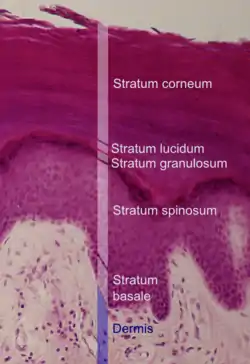
Histologic image of the epidermis with its layers named in white text.
It is named after Marcello Malpighi.
Basal cell carcinoma originates from the basal layer of the rete malpighii of the skin.
This layer is where almost all of the mitotic activity in epidermis occurs. The activity of these cells is increased by IK-1 (interleukin 1) and Epidermal growth factor. The activity is decreased by transforming growth factor. [5]
See also
References
- McGrath, J.A.; Eady, R.A.; Pope, F.M. (2004). Rook's Textbook of Dermatology (Seventh Edition). Blackwell Publishing. Pages 3.1-3.6. ISBN 978-0-632-06429-8.
- TheFreeDictionary > Malpighian layer Citing: *Saunders Comprehensive Veterinary Dictionary, 3 ed. 2007
- TheFreeDictionary > Malpighian layer Citing: *The American Heritage Medical Dictionary 2007
- Wilkinson, P.F. Millington, R. (2009). Skin (Digitally printed version ed.). Cambridge: Cambridge University Press. p. 49. ISBN 978-0-521-10681-8.
- Mescher, A. L., Mescher, A. L., & Junqueira, L. C. U. (2016). Junqueira's basic histology: Text and atlas (Fourteenth edition.). New York: McGraw-Hill Education.
This article is issued from Wikipedia. The text is licensed under Creative Commons - Attribution - Sharealike. Additional terms may apply for the media files.