Mycena haematopus
Mycena haematopus, commonly known as the bleeding fairy helmet, the burgundydrop bonnet, or the bleeding Mycena, is a species of fungus in the family Mycenaceae, of the order Agaricales. It is widespread and common in Europe and North America, and has also been collected in Japan and Venezuela. It is saprotrophic—meaning that it obtains nutrients by consuming decomposing organic matter—and the fruit bodies appear in small groups or clusters on the decaying logs, trunks, and stumps of deciduous trees, particularly beech. The fungus, first described scientifically in 1799, is classified in the section Lactipedes of the genus Mycena, along with other species that produce a milky or colored latex.
| Mycena haematopus | |
|---|---|
 | |
| Scientific classification | |
| Kingdom: | |
| Division: | |
| Class: | |
| Order: | |
| Family: | |
| Genus: | |
| Species: | M. haematopus |
| Binomial name | |
| Mycena haematopus | |
| Synonyms[1] | |
| Mycena haematopus | |
|---|---|
float | |
| gills on hymenium | |
| cap is conical or campanulate | |
| hymenium is adnate | |
| stipe is bare | |
| spore print is white | |
| ecology is saprotrophic | |
| edibility: not recommended | |
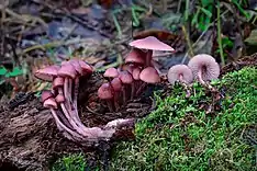
The fruit bodies of M. haematopus have caps that are up to 4 cm (1.6 in) wide, whitish gills, and a thin, fragile reddish-brown stem with thick coarse hairs at the base. They are characterized by their reddish color, the scalloped cap edges, and the dark red latex they "bleed" when cut or broken. Both the fruit bodies and the mycelia are weakly bioluminescent. M. haematopus produces various alkaloid pigments unique to this species. The edibility of the fruit bodies is not known definitively.
Taxonomy and naming
The species was initially named Agaricus haematopus by Christian Hendrik Persoon in 1799,[2] and later sanctioned under this name by Elias Magnus Fries in his 1821 Systema Mycologicum.[3] In the classification of Fries, only a few genera were named, and most agaric mushrooms were grouped in Agaricus, which was organized into a large number of tribes. Mycena haematopus gained its current name in 1871 when the German fungal taxonomist Paul Kummer raised many of Fries' Agaricus tribes to the level of genus, including Mycena.[4] In 1909 Franklin Sumner Earle placed the species in Galactopus,[5] a genus that is no longer considered separate from Mycena.[6] Mycena haematopus is placed in the section Lactipedes, a grouping of Mycenas characterized by the presence of a milky or colored latex in the stem and flesh of the cap.[7] The specific epithet is derived from Ancient Greek roots meaning "blood" (αἱματο-, haimato-) and "foot" (πους, pous).[8] It is commonly known as the blood-foot mushroom, the bleeding fairy helmet,[9] the burgundydrop bonnet,[10] or the bleeding Mycena.[11]
In 1914, Jakob Emanuel Lange described the variety M. haematopus var. marginata, characterized by the reddish color on the edge of the gills;[12] Mycena specialist Rudolph Arnold Maas Geesteranus considered the coloration of the gill edge too variable to have taxonomical significance.[13] Mycena haematopus var. cuspidata was initially found in Colorado in 1976, and described as a new variety by American mycologists Duane Mitchel and Alexander H. Smith two years later. The fruit bodies are characterized by a "beak" on the cap that often splits or collapses as the cap matures.[14] It was treated as Mycena sanguinolenta var. cuspidata by Maas Geesteranus in 1988.[13]
Description
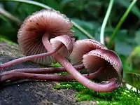
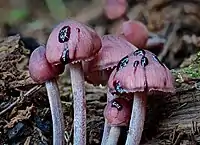
The fruit bodies of Mycena haematopus are the reproductive structures produced by cellular threads or hyphae which grow in rotting wood. The shape of the cap of the fruit body will vary depending on its maturity. Young caps, or "buttons", are ovoid (egg-shaped) to conical; later they are campanulate (bell-shaped), and as the fruit body matures, the margins (cap edge) lift upward so that the cap becomes somewhat flat with an umbo (a central nipple-shaped bump).[15] The fully grown cap can reach up to 4 cm (1.6 in) in diameter. The surface of the cap initially appears dry and covered with what appears to be a very fine whitish powder, but it soon becomes polished and moist. Mature caps appear somewhat translucent, and develop radial grooves mirroring the position of the gills underneath.[16] The color of the cap is reddish- or pinkish-brown, often tinged with violet, and paler towards the edge. The margin is wavy like the edge of a scallop, and may appear ragged because of lingering remnants of the partial veil.
The mushroom flesh can range from pale to the color of red wine (vinaceous), and has no distinctive odor. It oozes a red latex when cut.[11] The gills have an adnate attachment to the stem, meaning they are more or less directly attached to it. They are initially whitish or "grayish vinaceous" in color, and can develop reddish-brown stains. Between 20 and 30 gills reach from the cap edge to the stem, resulting in a gill spacing that is described as "close to subdistant"—gaps are visible between adjacent gills. There are additional gills, called lamellulae, that do not extend directly from the margin to the stem; these are arranged in two or three series (tiers) of equal length. The stem is up to 9 cm (3.5 in) tall and 0.1 to 0.2 cm (0.04 to 0.08 in) thick, hollow and brittle, and a dark reddish-brown color. In young fruit bodies, the upper part of the stem is densely covered with a pale cinnamon-colored powder which wears off with age. The stem has a mass of coarse hairs at the base. Like the cap, the stem also bleeds a red latex when it is cut or broken.[15][16]
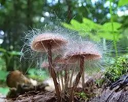
Mycena haematopus can be parasitized by Spinellus fusiger, another fungal species which gives the mushroom a strikingly hairy appearance.[8][17]
Microscopic characteristics
The spore print is white. The spores are elliptical, smooth, with dimensions of 8–11 by 5–7 µm. They are amyloid, meaning they will absorb iodine when stained with Melzer's reagent.[17] The spore-bearing cells (basidia) are 4-spored. Sterile cells called cystidia are numerous on the edges on the gills; they measure 33–60 µm (sometimes up to 80) by 9–12 µm. Cystidia that are present on the stipe (caulocystidia) appear in clusters, and clublike to irregular in shape, measuring 20–55 by 3.5–12.5 µm.[18] The gill tissue contains numerous lactifers, cells that produce the latex that is secreted when it is cut.[16]
The surface mycelium of M. haematopus is whitish and fluffy. Swelling at the terminal tips of hyphae (diameter up to 12 µm) is present, but not very abundant, and moniliform hyphae are very rare. Bioluminescence is present, but weak. Extracellular oxidase enzymes are present, consistent with its ecological role as a saprobe.[19]
Edibility
Although some sources claim that M. haematopus is edible,[20][21] it is "hardly worth collecting because of its small size."[11] Other sources consider the species inedible,[22] or recommend avoiding consumption, "since most of them have not yet been tested for toxins."[8] The taste of the mushroom is mild to slightly bitter.[23]
Similar species
Another Mycena that produces a reddish latex is Mycena sanguinolenta, the "terrestrial bleeding Mycena". It may be distinguished from M. haematopus in several ways: it is smaller, with cap diameters between 0.3 to 1 cm (0.1 to 0.4 in) wide; grows in groups rather than clusters; is found on leaves, dead branches, moss beds and pine needle beds rather than decaying wood; and the edges of its gills are consistently dark brownish-red.[24] Furthermore, range of cap color in M. sanguinolenta is different than in M. haematopus, varying from reddish-to orange-brown, and it lacks a band of partial veil remnants hanging from the margin.[21]
Ecology, distribution and habitat
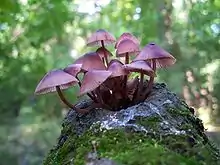
Mycena haematopus obtains nutrients from decomposing organic matter (saprobic) and the fruit bodies can typically be found growing on stumps and well-decayed logs, usually in groups that are joined together by a common base. The decomposition of woody debris on the forest floor is the result of the combined activity of a community of fungal species. In the sequential succession of mushrooms species, M. haematopus is a "late colonizer" fungus: its fruit bodies appear after the wood has first been decayed by white rot species. The initial stage of wood decay by white rot fungi involves the breakdown of "acid-unhydrolyzable residue" and holocellulose (a mixture of cellulose and hemicellulose).[25]
In North America, Mycena haematopus is known to be distributed from Alaska southward.[15] According to Mycena specialist Alexander H. Smith, it is "the commonest and the most easily recognized one in the genus."[16] The species is common in Europe,[11] and it has also been collected from Japan,[26] and Mérida, Venezuela, as the variety M. haematopus var. marginata.[27] In the Netherlands, M. haematopus is one of many mushrooms that can regularly be found fruiting on ancient timber wharves.[28] The fruit bodies can be found year-round in mild weather.[29]
Bioluminescence
Both the mycelia[19] and the fruit bodies of M. haematopus (both young and mature specimens) are reported to be bioluminescent. However, the luminescence is quite weak, and not visible to the dark-adapted eye; in one study, light emission was detectable only after 20 hours of exposure to X-ray film.[30] Although the biochemical basis of bioluminescence in M. haematopus has not been scientifically investigated, in general, bioluminescence is caused by the action of luciferases, enzymes that produce light by the oxidation of a luciferin (a pigment).[31] The biological purpose of bioluminescence in fungi is not definitively known, although several hypotheses have been suggested: it may help attract insects to help with spore dispersal,[32] it may be a by-product of other biochemical functions,[33] or it may help deter heterotrophs that might consume the fungus.[32]
Natural products

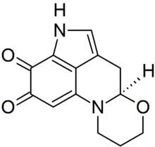
Several unique chemicals are produced by Mycena haematopus. The primary pigment is haematopodin B, which is so chemically sensitive (breaking down upon exposure to air and light) that its more stable breakdown product, haematopodin,[34] was known before its eventual discovery and characterization in 2008.[35] A chemical synthesis for haematopodin was reported in 1996.[36] Haematopodins are the first pyrroloquinoline alkaloids discovered in fungi; pyrroloquinolines combine the structures of pyrrole and quinoline, both heterocyclic aromatic organic compounds. Compounds of this type also occur in marine sponges and are attracting research interest due to various biological properties, such as cytotoxicity against tumor cell lines, and both antifungal and antimicrobial activities.[34] Additional alkaloid compounds in M. haematopus include the red pigments mycenarubins D, E and F. Prior to the discovery of these compounds, pyrroloquinoline alkaloids were considered to be rare in terrestrial sources.[35]
See also
References
- "Species Fungorum – Species synonymy". Index Fungorum. CAB International. Archived from the original on 2011-06-10. Retrieved 2010-04-25.
- Persoon CH. (1799). Observationes mycologicae (in Latin). 2. Leipzig, Germany: Gesnerus, Usterius & Wolfius. p. 56.
- Fries EM. (1821). Systema Mycologicum (in Latin). 1. Lundae: Ex Officina Berlingiana. p. 149.
- Kummer P. (1871). Der Führer in die Pilzkunde (in German). Zerbst. p. 109.
- Earle FS. (1906). "The genera of North American gill fungi". Bulletin of the New York Botanical Garden. 5: 373–451.
- Kirk PM, Cannon PF, Minter DW, Stalpers JA (2008). Dictionary of the Fungi (10th ed.). Wallingford: CAB International. p. 270. ISBN 978-0-85199-826-8.
- Smith, 1947, p. 132.
- Volk T. (June 2002). "Mycena haematopus, the blood-foot mushroom". Tom Volk's Fungus of the Month. UW-Madison Department of Botany. Retrieved 2010-02-26.
- Sept JD. (2006). Common Mushrooms of the Northwest: Alaska, Western Canada & the Northwestern United States. Sechelt, BC, Canada: Calypso Publishing. p. 32. ISBN 978-0-9739819-0-2.
- Marren P (2009-10-29). "The magic of Britain's wild mushrooms". Telegraph.co.uk. Retrieved 2010-03-01.
- Hall IR. (2003). Edible and Poisonous Mushrooms of the World. Portland: Timber Press. p. 164. ISBN 978-0-88192-586-9.
- Lange JE. (1914). "Studies in the Agarics of Denmark. Part I. Mycena". Dansk Botanisk Arkiv. 1 (5): 1–40.
- Maas Geesteranus RA. (1988). "Conspectus of the Mycenas of the northern hemisphere 10. Sections Lactipedes, Sanguinolentae, Galactopoda, and Crocatae". Proceedings of the Koninklijke Nederlandse Akademie van Wetenschappen Series C Biological and Medical Sciences. 91 (4): 377–404. ISSN 0023-3374.
- Mitchel DH, Smith AH (1978). "Notes on Colorado fungi III: New and interesting mushrooms from the aspen zone". Mycologia. 70 (5): 1040–63. doi:10.2307/3759137. JSTOR 3759137.
- Orr DB, Orr RT (1979). Mushrooms of Western North America. Berkeley: University of California Press. p. 235. ISBN 978-0-520-03656-7.
- Smith, 1947, pp. 140–44.
- Healy RA, Huffman DR, Tiffany LH, Knaphaus G (2008). Mushrooms and Other Fungi of the Midcontinental United States (Bur Oak Guide). Iowa City: University of Iowa Press. p. 147. ISBN 978-1-58729-627-7.
- Aronsen A. "Mycena haematopus (Pers.) P. Kumm". A key to the Mycenas of Norway. Archived from the original on 2010-10-12. Retrieved 2010-03-01.
- Treu R, Agerer R (1990). "Culture characteristics of some Mycena species". Mycotaxon. 38: 279–309.
- McKnight VB, McKnight KH (1987). A Field Guide to Mushrooms: North America. Boston: Houghton Mifflin. Plate 19. ISBN 978-0-395-91090-0.
- Arora D. (1986). Mushrooms Demystified: A Comprehensive Guide to the Fleshy Fungi. Berkeley: Ten Speed Press. p. 231. ISBN 978-0-89815-169-5.
- Jordan M. (2004). The Encyclopedia of Fungi of Britain and Europe. London: Frances Lincoln. p. 168. ISBN 978-0-7112-2378-3.
- Kuo M (December 2010). "Mycena haematopus". MushroomExpert.Com. Retrieved 2013-06-15.
- Smith, 1947, pp. 146–49.
- Fukasawa Y, Osono T, Takeda H (2008). "Dynamics of physicochemical properties and occurrence of fungal fruit bodies during decomposition of coarse woody debris of Fagus crenata". Journal of Forest Research. 14: 20–29. doi:10.1007/s10310-008-0098-0. S2CID 39919592.
- Desjardin DE, Oliveira AG, Stevani CV (2008). "Fungi bioluminescence revisited". Photochemical & Photobiological Sciences. 7 (2): 170–82. doi:10.1039/b713328f. PMID 18264584.
- Dennis RWG. (1961). "Fungi venezuelani: IV. Agaricales". Kew Bulletin. 15 (1): 67–156. doi:10.2307/4115784. JSTOR 4115784.
- De Held-Jager CM. (1979). "The mushrooms of timber wharves in the vicinity of Gouda Netherlands". Coolia. 22 (2): 52–56. ISSN 0929-7839.
- Bessette A, Bessette AR, Fischer DW (1997). Mushrooms of Northeastern North America. Hong Kong: Syracuse University Press. pp. 211–12. ISBN 978-0-8156-0388-7.
- Bermudes D, Petersen RH, Nealson KH (1992). "Low-level bioluminescence detected in Mycena haematopus basidiocarps" (PDF). Mycologia. 84 (5): 799–802. doi:10.2307/3760392. hdl:10211.2/2130. JSTOR 3760392.
- Shimomura O. (2006). Bioluminescence: Chemical Principles and Methods. Singapore: World Scientific. pp. 268–75. ISBN 978-981-256-801-4.
- Sivinski J. (1981). "Arthropods attracted to luminous fungi". Psyche. 88 (3–4): 383–90. doi:10.1155/1981/79890.
- Buller AHR. (1924). "The bioluminescence of Panus stypticus". Researches on Fungi. III. London: Longsman, Green and Co. pp. 357–431.
- Baumann C, Bröckelmann M, Fugmann B, Steffan B, Steglich W, Sheldrick WS (1993). "Pigments of fungi. 62. Haematopodin, an unusual pyrroloquinoline derivative isolated from the fungus Mycena haematopus, Agaricales". Angewandte Chemie International Edition in English. 32 (7): 1087–89. doi:10.1002/anie.199310871.
- Peters S, Jaeger RJR, Spiteller P (2008). "Red pyrroloquinoline alkaloids from the mushroom Mycena haematopus". European Journal of Organic Chemistry. 2008 (2): 319–23. doi:10.1002/ejoc.200700739.
- Hopmann C, Steglich W (1996). "Synthesis of haematopodin – A pigment from the mushroom Mycena haematopus (Basidiomycetes)". Liebigs Annalen. 1996 (7): 1117–20. doi:10.1002/jlac.199619960709.
Books cited
- Smith AH. (1947). North American species of Mycena. Ann Arbor: University of Michigan Press.
External links
| Wikimedia Commons has media related to Mycena haematopus. |
- Fungi Growing on Wood by Gary Emberger
- California Fungi Michael Wood and Fred Stevens
