Pinocytosis
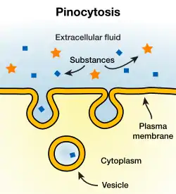
In cellular biology, pinocytosis, otherwise known as fluid endocytosis and bulk-phase pinocytosis, is a mode of endocytosis in which small particles suspended in extracellular fluid are brought into the cell through an invagination of the cell membrane, resulting in a suspension of the particles within a small vesicle inside the cell. These pinocytotic vesicles then typically fuse with early endosomes to hydrolyze (break down) the particles.
Pinocytosis is further segregated into the pathways macropinocytosis, clathrin-mediated endocytosis, caveolin-mediated endocytosis, or clathrin- and caveolin-independent endocytosis, all of which differ by the mechanism of vesicle formation as well as the resulting size of these vesicles.
Pinocytosis is variably subdivided into categories depending on molecular mechanism and the fate of the internalized molecules. Pinocytosis is, in some cases, considered to be a constitutive process, while in others it is receptor-mediated and highly regulated.
Pinocytosis
In humans, this process occurs primarily for absorption of fat droplets. In endocytosis the cell plasma membrane extends and folds around desired extracellular material, forming a pouch that pinches off creating an internalized vesicle. The invaginated pinocytosis vesicles are much smaller than those generated by phagocytosis. The vesicles eventually fuse with the lysosome whereupon the vesicle contents are digested. Pinocytosis involves a considerable investment of cellular energy in the form of ATP
2 Pinocytosis and ATP
Pinocytosis is used primarily for clearing extracellular fluids (ECF) and as part of immune surveillance.[1] In contrast to phagocytosis, it generates very small amounts of ATP from the wastes of alternative substances such as lipids (fat). Unlike receptor-mediated endocytosis, pinocytosis is nonspecific in the substances that it transports. The cell takes in surrounding fluids, including all solutes present. Pinocytosis also works as phagocytosis; the only difference is that phagocytosis is specific in the substances it transports. Phagocytosis engulfs whole particles, which are later broken down by enzymes, such as cathepsins, and absorbed into the cells. Pinocytosis, on the other hand, is when the cell engulfs already-dissolved or broken-down food.
Pinocytosis is non-specific and non-absorptive. Molecule-specific endocytosis is called receptor-mediated endocytosis.
Etymology and pronunciation
The word pinocytosis (/ˌpɪnəsaɪˈtoʊsɪs, ˌpaɪ-, -noʊ-, -sə-/[2][3][4]) uses combining forms of pino- + cyto- + -osis, all New Latin from Greek, reflecting píno, to drink, and cytosis. The term was proposed by W. H. Lewis in 1931.[5]
Non-specific, adsorptive pinocytosis
Non-specific, adsorptive pinocytosis is a form of endocytosis, a process in which small particles are taken in by a cell by splitting off small vesicles from the cell surface.[6] Cationic proteins bind to the negative cell surface and are taken up via the clathrin-mediated system, thus the uptake is intermediate between receptor-mediated endocytosis and non-specific, non-adsorptive pinocytosis. The clathrin-coated pits occupy about 2% of the surface area of the cell and only last about a minute, with an estimated 2500 leaving the average cell surface each minute. The clathrin coats are lost almost immediately, and the membrane is subsequently recycled to the cell surface.
Macropinocytosis
Macropinocytosis is a clathrin-independent endocytic mechanism that can be activated in practically all animal cells. In most cell types, it does not occur continuously but rather is induced for a limited time in response to cell-surface receptor activation by specific cargoes, including growth factors, ligands of integrins, apoptotic cell remnants, and some viruses. These ligands activate a complex signaling pathway, resulting in a change in actin dynamics and the formation of cell-surface protrusions, called ruffles. When ruffles collapse back onto the membrane, large fluid-filled endocytic vesicles form, called macropinosomes which can transiently increase the bulk fluid uptake of a cell by up to tenfold. Macropinocytosis is a solely degradative pathway: macropinosomes acidify and then fuse with late endosomes or endolysosomes, without recycling their cargo back to the plasma membrane.[7]
References
- Abbas, Abul, et al. "Basic Immunology: Functions and Disorders of the Immune System." 5th ed. Elsevier, 2016. p.69
- "Pinocytosis". Oxford Dictionaries UK Dictionary. Oxford University Press. Retrieved 2016-01-22.
- "Pinocytosis". Merriam-Webster Dictionary. Retrieved 2016-01-22.
- "Pinocytosis". Dictionary.com Unabridged. Random House. Retrieved 2016-01-22.
- Rieger, R.; Michaelis, A.; Green, M.M. 1991. Glossary of Genetics. Classical and Molecular (Fifth edition). Springer-Verlag, Berlin, .
- Alberts, Johnson, Lewis, Raff, Roberts, Walter: "Molecular Biology of the Cell", Fourth Edition, Copyright 2002 P.748
- Alberts, Bruce (2015). Molecular biology of the cell (Sixth ed.). New York, NY. p. 732. ISBN 978-0-8153-4432-2. OCLC 887605755.
- Campbell, Reece, Mitchell: "Biology", Sixth Edition, Copyright 2002 P. 151
- Marshall, Ben, Incredible Biological Advancements of the 20th Century, Copyright 2001 p. 899
- Alrt, Pablo, Global Society Harvard study, copyright 2003 p. 189
- Brooker, Robert: "Biology", Second Edition, Copyright 2011 p. 116
- Cherrr, Malik, The Only Edition, Copyright 2012, p. 256
- Abbas, Abul, et al. "Basic Immunology: Functions and Disorders of the Immune System." 5th ed. Elsevier, 2016. p.69