Retromandibular vein
The retromandibular vein (temporomaxillary vein, posterior facial vein) is a major vein of the face.
| Posterior facial vein | |
|---|---|
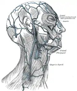 Veins of the head and neck (retromandibular vein visible at center). | |
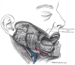 Dissection, showing salivary glands of right side (retromandibular vein visible at bottom center). | |
| Details | |
| Source | Superficial temporal vein, maxillary veins |
| Drains to | External jugular vein |
| Artery | Facial artery |
| Identifiers | |
| Latin | Vena retromandibularis, vena facialis posterior |
| TA98 | A12.3.05.031 |
| TA2 | 4831 |
| FMA | 50928 |
| Anatomical terminology | |
Structure
The retromandibular vein is formed by the union of the superficial temporal and maxillary veins.[1][2] It descends in the substance of the parotid gland, superficial to the external carotid artery (but beneath the facial nerve),[3] between the ramus of the mandible and the sternocleidomastoideus muscle.
It divides into two branches:
- an anterior, which passes forward and joins anterior facial vein, to form the common facial vein, which then drains into the internal jugular vein.[4]
- a posterior, which is joined by the posterior auricular vein and becomes the external jugular vein.[4][5]
Function
The retromandibular vein provides venous drainage to the superior cranium, and significant drainage to the ear.[6]
Clinical significance
Parrot's sign is a sensation of pain when pressure is applied to the retromandibular region.
Additional images
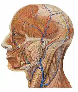 Lateral head anatomy detail
References
This article incorporates text in the public domain from page 646 of the 20th edition of Gray's Anatomy (1918)
- Thompson, Stevan H.; Yeung, Alison Y. (2016-01-01), Hupp, James R.; Ferneini, Elie M. (eds.), "4 - Anatomy Relevant to Head, Neck, and Orofacial Infections", Head, Neck, and Orofacial Infections, St. Louis: Elsevier, pp. 60–93, doi:10.1016/b978-0-323-28945-0.00004-1, ISBN 978-0-323-28945-0, retrieved 2020-11-11
- Cunningham, Larry L.; Card, Aaron Sterling (2012-01-01), Bagheri, Shahrokh C.; Bell, R. Bryan; Khan, Husain Ali (eds.), "Chapter 38 - Mandibular Subcondylar Fractures", Current Therapy In Oral and Maxillofacial Surgery, Saint Louis: W.B. Saunders, pp. 298–304, doi:10.1016/b978-1-4160-2527-6.00038-4, ISBN 978-1-4160-2527-6, retrieved 2020-11-11
- Loukota, Richard A.; Abdel-Galil, Khalid (2017-01-01), Brennan, Peter A.; Schliephake, Henning; Ghali, G. E.; Cascarini, Luke (eds.), "6 - Condylar Fractures", Maxillofacial Surgery (Third Edition), Churchill Livingstone, pp. 74–92, doi:10.1016/b978-0-7020-6056-4.00006-x, ISBN 978-0-7020-6056-4, retrieved 2020-11-11
- Cramer, Gregory D. (2014-01-01), Cramer, Gregory D.; Darby, Susan A. (eds.), "Chapter 5 - The Cervical Region", Clinical Anatomy of the Spine, Spinal Cord, and Ans (Third Edition), Saint Louis: Mosby, pp. 135–209, doi:10.1016/b978-0-323-07954-9.00005-0, ISBN 978-0-323-07954-9, retrieved 2020-11-11
- Drake, Richard L. (Richard Lee), 1950- (2005). Gray's anatomy for students. Vogl, Wayne., Mitchell, Adam W. M., Gray, Henry, 1825-1861. Philadelphia: Elsevier/Churchill Livingstone. ISBN 0-443-06612-4. OCLC 55139039.CS1 maint: multiple names: authors list (link)
- Posnick, Jeffrey C. (2014-01-01), Posnick, Jeffrey C. (ed.), "39 - Aesthetic Alteration of Prominent Ears: Evaluation and Surgery", Orthognathic Surgery, St. Louis: W.B. Saunders, pp. 1703–1745, doi:10.1016/b978-1-4557-2698-1.00039-3, ISBN 978-1-4557-2698-1, retrieved 2020-11-11
External links
- Anatomy photo:27:13-0103 at the SUNY Downstate Medical Center - "Infratemporal fossa: The Pterygoid plexus of Veins"
- lesson4 at The Anatomy Lesson by Wesley Norman (Georgetown University) (parotid2)
- Tufts.edu
| Authority control |
|---|