Thioredoxin
Thioredoxin is a class of small redox proteins known to be present in all organisms. It plays a role in many important biological processes, including redox signaling. In humans, thioredoxins are encoded by TXN and TXN2 genes.[5][6] Loss-of-function mutation of either of the two human thioredoxin genes is lethal at the four-cell stage of the developing embryo. Although not entirely understood, thioredoxin plays a central role in humans and is increasingly linked to medicine through their response to reactive oxygen species (ROS). In plants, thioredoxins regulate a spectrum of critical functions, ranging from photosynthesis to growth, flowering and the development and germination of seeds. They have also recently been found to play a role in cell-to-cell communication.[7]
Function
Thioredoxins are proteins that act as antioxidants by facilitating the reduction of other proteins by cysteine thiol-disulfide exchange. Thioredoxins are found in nearly all known organisms and are essential for life in mammals.[8][9]
Thioredoxin is a 12-kD oxidoreductase enzyme containing a dithiol-disulfide active site. It is ubiquitous and found in many organisms from plants and bacteria to mammals. Multiple in vitro substrates for thioredoxin have been identified, including ribonuclease, choriogonadotropins, coagulation factors, glucocorticoid receptor, and insulin. Reduction of insulin is classically used as an activity test.[10]
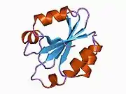
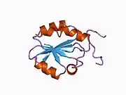
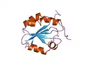
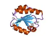
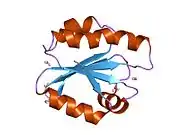
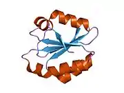
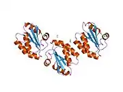
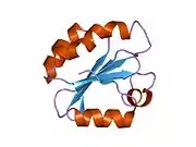
Thioredoxins are characterized at the level of their amino acid sequence by the presence of two vicinal cysteines in a CXXC motif. These two cysteines are the key to the ability of thioredoxin to reduce other proteins. Thioredoxin proteins also have a characteristic tertiary structure termed the thioredoxin fold.
The thioredoxins are kept in the reduced state by the flavoenzyme thioredoxin reductase, in a NADPH-dependent reaction.[11] Thioredoxins act as electron donors to peroxidases and ribonucleotide reductase.[12] The related glutaredoxins share many of the functions of thioredoxins, but are reduced by glutathione rather than a specific reductase.
The benefit of thioredoxins to reduce oxidative stress is shown by transgenic mice that overexpress thioredoxin, are more resistant to inflammation, and live 35% longer[13] — supporting the free radical theory of aging. However, the controls of this study were short lived, which may have contributed to the apparent increase in longevity.[14] Trx1 can regulate non-redox post-translational modifications.[15] In the mice with cardiac-specific overexpression of Trx1, the proteomics study found that SET and MYND domain-containing protein 1 (SMYD1), a lysine methyltransferase highly expressed in cardiac and other muscle tissues, is also upregulated. This suggests that Trx1 may also play an role in protein methylation via regulating SMYD1 expression, which is independent of its oxidoreductase activity.[15]
Plants have an unusually complex complement of Trxs composed of six well-defined types (Trxs f, m, x, y, h, and o) that reside in different cell compartments and function in an array of processes. In 2010 it was discovered for the first time that thioredoxin proteins are able to move from cell to cell, representing a novel form of cellular communication in plants.[7]
Mechanism of action
The primary function of Thioredoxin (Trx) is the reduction of oxidized cysteine residues and the cleavage of disulfide bonds.[16] For Trx1, this process begins by attack of Cys32, one of the residues conserved in the thioredoxin CXXC motif, onto the oxidized group of the substrate.[17] Almost immediately after this event Cys35, the other conserved Cys residue in Trx1, forms a disulfide bond with Cys32, thereby transferring 2 electrons to the substrate which is now in its reduced form. Oxidized Trx1 is then reduced by thioredoxin reductase, which in turn is reduced by NADPH as described above.[17]

Interactions
Thioredoxin has been shown to interact with:
- ASK1,[18][19][20]
- Collagen, type I, alpha 1,[21]
- Glucocorticoid receptor,[22]
- SENP1,[23]
- TXNIP.[24]
- NF-κB – by reducing a disulfide bond in NF-κB, Trx1 promotes binding of this transcription factor to DNA.[25]
- AP1 via Ref1 – Trx1 indirectly increases the DNA-binding activity of activator protein 1 (AP1) by reducing the DNA repair enzyme redox factor 1 (Ref-1), which in turn reduces AP1 in an example of a redox regulation cascade.[26]
- AMPK – AMPK function in cardiomyocytes is preserved during oxidative stress due to an interaction between AMPK and Trx1. By forming a disulfide bridge between the two proteins, Trx1 prevents the formation and aggregation of oxidized AMPK, thereby allowing AMPK to function normally and participate in signaling cascades.[27]
Effect on cardiac hypertrophy
Trx1 has been shown to downregulate cardiac hypertrophy, the thickening of the walls of the lower heart chambers, by interactions with several different targets. Trx1 upregulates the transcriptional activity of nuclear respiratory factors 1 and 2 (NRF1 and NRF2) and stimulates the expression of peroxisome proliferator-activated receptor γ coactivator 1-α (PGC-1α).[28][29] Furthermore, Trx1 reduces two cysteine residues in histone deacetylase 4 (HDAC4), which allows HDAC4 to be imported from the cytosol, where the oxidized form resides,[30] into the nucleus.[31] Once in the nucleus, reduced HDAC4 downregulates the activity of transcription factors such as NFAT that mediate cardiac hypertrophy.[17] Trx 1 also controls microRNA levels in the heart and has been found to inhibit cardiac hypertrophy by upregulating miR-98/let-7.[32] Trx1 can regulate the expression level of SMYD1, thus may indirectly modulate protein methylation for purpose of cardiac protection.[15]
Thioredoxin in skin care
Thioredoxin is used in skin care products as an antioxidant in conjunction with glutaredoxin and glutathione.
See also
- RuBisCO - enzyme activity regulated by thioredoxin
- Peroxiredoxin - enzyme activity regulated by thioredoxin
- Thioredoxin fold
References
- GRCh38: Ensembl release 89: ENSG00000136810 - Ensembl, May 2017
- GRCm38: Ensembl release 89: ENSMUSG00000028367 - Ensembl, May 2017
- "Human PubMed Reference:". National Center for Biotechnology Information, U.S. National Library of Medicine.
- "Mouse PubMed Reference:". National Center for Biotechnology Information, U.S. National Library of Medicine.
- Wollman EE, d'Auriol L, Rimsky L, Shaw A, Jacquot JP, Wingfield P, Graber P, Dessarps F, Robin P, Galibert F (October 1988). "Cloning and expression of a cDNA for human thioredoxin". The Journal of Biological Chemistry. 263 (30): 15506–12. PMID 3170595.
- "Entrez Gene: TXN2 thioredoxin 2".
- Meng L, Wong JH, Feldman LJ, Lemaux PG, Buchanan BB (February 2010). "A membrane-associated thioredoxin required for plant growth moves from cell to cell, suggestive of a role in intercellular communication". Proceedings of the National Academy of Sciences of the United States of America. 107 (8): 3900–5. doi:10.1073/pnas.0913759107. PMC 2840455. PMID 20133584.
- Holmgren A (August 1989). "Thioredoxin and glutaredoxin systems" (PDF). The Journal of Biological Chemistry. 264 (24): 13963–6. PMID 2668278.
- Nordberg J, Arnér ES (December 2001). "Reactive oxygen species, antioxidants, and the mammalian thioredoxin system". Free Radical Biology & Medicine. 31 (11): 1287–312. doi:10.1016/S0891-5849(01)00724-9. PMID 11728801.
- "Entrez Gene: TXN thioredoxin".
- Mustacich D, Powis G (February 2000). "Thioredoxin reductase". The Biochemical Journal. 346 (1): 1–8. doi:10.1042/0264-6021:3460001. PMC 1220815. PMID 10657232.
- Arnér ES, Holmgren A (October 2000). "Physiological functions of thioredoxin and thioredoxin reductase". European Journal of Biochemistry. 267 (20): 6102–9. doi:10.1046/j.1432-1327.2000.01701.x. PMID 11012661.
- Yoshida T, Nakamura H, Masutani H, Yodoi J (December 2005). "The involvement of thioredoxin and thioredoxin binding protein-2 on cellular proliferation and aging process". Annals of the New York Academy of Sciences. 1055: 1–12. doi:10.1196/annals.1323.002. PMID 16387713. S2CID 37043674.
- Muller FL, Lustgarten MS, Jang Y, Richardson A, Van Remmen H (August 2007). "Trends in oxidative aging theories". Free Radical Biology & Medicine. 43 (4): 477–503. doi:10.1016/j.freeradbiomed.2007.03.034. PMID 17640558.
- Liu T, Wu C, Jain MR, Nagarajan N, Yan L, Dai H, Cui C, Baykal A, Pan S, Ago T, Sadoshima J, Li H (December 2015). "Master redox regulator Trx1 upregulates SMYD1 & modulates lysine methylation". Biochimica et Biophysica Acta (BBA) - Proteins and Proteomics. 1854 (12): 1816–1822. doi:10.1016/j.bbapap.2015.09.006. PMC 4721509. PMID 26410624.
- Nakamura H, Nakamura K, Yodoi J (1997-01-01). "Redox regulation of cellular activation". Annual Review of Immunology. 15 (1): 351–69. doi:10.1146/annurev.immunol.15.1.351. PMID 9143692.
- Nagarajan N, Oka S, Sadoshima J (December 2016). "Modulation of signaling mechanisms in the heart by thioredoxin 1". Free Radical Biology & Medicine. 109: 125–131. doi:10.1016/j.freeradbiomed.2016.12.020. PMC 5462876. PMID 27993729.
- Liu Y, Min W (June 2002). "Thioredoxin promotes ASK1 ubiquitination and degradation to inhibit ASK1-mediated apoptosis in a redox activity-independent manner". Circulation Research. 90 (12): 1259–66. doi:10.1161/01.res.0000022160.64355.62. PMID 12089063.
- Morita K, Saitoh M, Tobiume K, Matsuura H, Enomoto S, Nishitoh H, Ichijo H (November 2001). "Negative feedback regulation of ASK1 by protein phosphatase 5 (PP5) in response to oxidative stress". The EMBO Journal. 20 (21): 6028–36. doi:10.1093/emboj/20.21.6028. PMC 125685. PMID 11689443.
- Saitoh M, Nishitoh H, Fujii M, Takeda K, Tobiume K, Sawada Y, Kawabata M, Miyazono K, Ichijo H (May 1998). "Mammalian thioredoxin is a direct inhibitor of apoptosis signal-regulating kinase (ASK) 1". The EMBO Journal. 17 (9): 2596–606. doi:10.1093/emboj/17.9.2596. PMC 1170601. PMID 9564042.
- Matsumoto K, Masutani H, Nishiyama A, Hashimoto S, Gon Y, Horie T, Yodoi J (July 2002). "C-propeptide region of human pro alpha 1 type 1 collagen interacts with thioredoxin". Biochemical and Biophysical Research Communications. 295 (3): 663–7. doi:10.1016/s0006-291x(02)00727-1. PMID 12099690.
- Makino Y, Yoshikawa N, Okamoto K, Hirota K, Yodoi J, Makino I, Tanaka H (January 1999). "Direct association with thioredoxin allows redox regulation of glucocorticoid receptor function". The Journal of Biological Chemistry. 274 (5): 3182–8. doi:10.1074/jbc.274.5.3182. PMID 9915858.
- Li X, Luo Y, Yu L, Lin Y, Luo D, Zhang H, He Y, Kim YO, Kim Y, Tang S, Min W (April 2008). "SENP1 mediates TNF-induced desumoylation and cytoplasmic translocation of HIPK1 to enhance ASK1-dependent apoptosis". Cell Death and Differentiation. 15 (4): 739–50. doi:10.1038/sj.cdd.4402303. PMID 18219322.
- Nishiyama A, Matsui M, Iwata S, Hirota K, Masutani H, Nakamura H, Takagi Y, Sono H, Gon Y, Yodoi J (July 1999). "Identification of thioredoxin-binding protein-2/vitamin D(3) up-regulated protein 1 as a negative regulator of thioredoxin function and expression". The Journal of Biological Chemistry. 274 (31): 21645–50. doi:10.1074/jbc.274.31.21645. PMID 10419473.
- Matthews JR, Wakasugi N, Virelizier JL, Yodoi J, Hay RT (August 1992). "Thioredoxin regulates the DNA binding activity of NF-kappa B by reduction of a disulphide bond involving cysteine 62". Nucleic Acids Research. 20 (15): 3821–30. doi:10.1093/nar/20.15.3821. PMC 334054. PMID 1508666.
- Hirota K, Matsui M, Iwata S, Nishiyama A, Mori K, Yodoi J (April 1997). "AP-1 transcriptional activity is regulated by a direct association between thioredoxin and Ref-1". Proceedings of the National Academy of Sciences of the United States of America. 94 (8): 3633–8. doi:10.1073/pnas.94.8.3633. PMC 20492. PMID 9108029.
- Shao D, Oka S, Liu T, Zhai P, Ago T, Sciarretta S, Li H, Sadoshima J (February 2014). "A redox-dependent mechanism for regulation of AMPK activation by Thioredoxin1 during energy starvation". Cell Metabolism. 19 (2): 232–45. doi:10.1016/j.cmet.2013.12.013. PMC 3937768. PMID 24506865.
- Ago T, Yeh I, Yamamoto M, Schinke-Braun M, Brown JA, Tian B, Sadoshima J (2006). "Thioredoxin1 upregulates mitochondrial proteins related to oxidative phosphorylation and TCA cycle in the heart". Antioxidants & Redox Signaling. 8 (9–10): 1635–50. doi:10.1089/ars.2006.8.1635. PMID 16987018.
- Yamamoto M, Yang G, Hong C, Liu J, Holle E, Yu X, Wagner T, Vatner SF, Sadoshima J (November 2003). "Inhibition of endogenous thioredoxin in the heart increases oxidative stress and cardiac hypertrophy". The Journal of Clinical Investigation. 112 (9): 1395–406. doi:10.1172/JCI17700. PMC 228400. PMID 14597765.
- Matsushima S, Kuroda J, Ago T, Zhai P, Park JY, Xie LH, Tian B, Sadoshima J (February 2013). "Increased oxidative stress in the nucleus caused by Nox4 mediates oxidation of HDAC4 and cardiac hypertrophy". Circulation Research. 112 (4): 651–63. doi:10.1161/CIRCRESAHA.112.279760. PMC 3574183. PMID 23271793.
- Ago T, Liu T, Zhai P, Chen W, Li H, Molkentin JD, Vatner SF, Sadoshima J (June 2008). "A redox-dependent pathway for regulating class II HDACs and cardiac hypertrophy". Cell. 133 (6): 978–93. doi:10.1016/j.cell.2008.04.041. PMID 18555775. S2CID 2678474.
- Yang Y, Ago T, Zhai P, Abdellatif M, Sadoshima J (February 2011). "Thioredoxin 1 negatively regulates angiotensin II-induced cardiac hypertrophy through upregulation of miR-98/let-7". Circulation Research. 108 (3): 305–13. doi:10.1161/CIRCRESAHA.110.228437. PMC 3249645. PMID 21183740.
Further reading
- Arnér ES, Holmgren A (October 2000). "Physiological functions of thioredoxin and thioredoxin reductase". European Journal of Biochemistry. 267 (20): 6102–9. doi:10.1046/j.1432-1327.2000.01701.x. PMID 11012661.
- Nishinaka Y, Masutani H, Nakamura H, Yodoi J (2002). "Regulatory roles of thioredoxin in oxidative stress-induced cellular responses". Redox Report. 6 (5): 289–95. doi:10.1179/135100001101536427. PMID 11778846. S2CID 34079507.
- Ago T, Sadoshima J (November 2006). "Thioredoxin and ventricular remodeling". Journal of Molecular and Cellular Cardiology. 41 (5): 762–73. doi:10.1016/j.yjmcc.2006.08.006. PMC 1852508. PMID 17007870.
- Tonissen KF, Wells JR (June 1991). "Isolation and characterization of human thioredoxin-encoding genes". Gene. 102 (2): 221–8. doi:10.1016/0378-1119(91)90081-L. PMID 1874447.
- Martin H, Dean M (February 1991). "Identification of a thioredoxin-related protein associated with plasma membranes". Biochemical and Biophysical Research Communications. 175 (1): 123–8. doi:10.1016/S0006-291X(05)81209-4. PMID 1998498.
- Forman-Kay JD, Clore GM, Wingfield PT, Gronenborn AM (March 1991). "High-resolution three-dimensional structure of reduced recombinant human thioredoxin in solution". Biochemistry. 30 (10): 2685–98. doi:10.1021/bi00224a017. PMID 2001356.
- Jacquot JP, de Lamotte F, Fontecave M, Schürmann P, Decottignies P, Miginiac-Maslow M, Wollman E (December 1990). "Human thioredoxin reactivity-structure/function relationship". Biochemical and Biophysical Research Communications. 173 (3): 1375–81. doi:10.1016/S0006-291X(05)80940-4. PMID 2176490.
- Forman-Kay JD, Clore GM, Driscoll PC, Wingfield P, Richards FM, Gronenborn AM (August 1989). "A proton nuclear magnetic resonance assignment and secondary structure determination of recombinant human thioredoxin". Biochemistry. 28 (17): 7088–97. doi:10.1021/bi00443a045. PMID 2684271.
- Tagaya Y, Maeda Y, Mitsui A, Kondo N, Matsui H, Hamuro J, Brown N, Arai K, Yokota T, Wakasugi H (March 1989). "ATL-derived factor (ADF), an IL-2 receptor/Tac inducer homologous to thioredoxin; possible involvement of dithiol-reduction in the IL-2 receptor induction". The EMBO Journal. 8 (3): 757–64. doi:10.1002/j.1460-2075.1989.tb03436.x. PMC 400872. PMID 2785919.
- Wollman EE, d'Auriol L, Rimsky L, Shaw A, Jacquot JP, Wingfield P, Graber P, Dessarps F, Robin P, Galibert F (October 1988). "Cloning and expression of a cDNA for human thioredoxin". The Journal of Biological Chemistry. 263 (30): 15506–12. PMID 3170595.
- Heppell-Parton A, Cahn A, Bench A, Lowe N, Lehrach H, Zehetner G, Rabbitts P (March 1995). "Thioredoxin, a mediator of growth inhibition, maps to 9q31". Genomics. 26 (2): 379–81. doi:10.1016/0888-7543(95)80223-9. PMID 7601465.
- Qin J, Clore GM, Kennedy WM, Huth JR, Gronenborn AM (March 1995). "Solution structure of human thioredoxin in a mixed disulfide intermediate complex with its target peptide from the transcription factor NF kappa B". Structure. 3 (3): 289–97. doi:10.1016/S0969-2126(01)00159-9. PMID 7788295.
- Kato S, Sekine S, Oh SW, Kim NS, Umezawa Y, Abe N, Yokoyama-Kobayashi M, Aoki T (December 1994). "Construction of a human full-length cDNA bank". Gene. 150 (2): 243–50. doi:10.1016/0378-1119(94)90433-2. PMID 7821789.
- Qin J, Clore GM, Gronenborn AM (June 1994). "The high-resolution three-dimensional solution structures of the oxidized and reduced states of human thioredoxin". Structure. 2 (6): 503–22. doi:10.1016/S0969-2126(00)00051-4. PMID 7922028.
- Gasdaska PY, Oblong JE, Cotgreave IA, Powis G (August 1994). "The predicted amino acid sequence of human thioredoxin is identical to that of the autocrine growth factor human adult T-cell derived factor (ADF): thioredoxin mRNA is elevated in some human tumors". Biochimica et Biophysica Acta (BBA) - Gene Structure and Expression. 1218 (3): 292–6. doi:10.1016/0167-4781(94)90180-5. PMID 8049254.
- Qin J, Clore GM, Kennedy WP, Kuszewski J, Gronenborn AM (May 1996). "The solution structure of human thioredoxin complexed with its target from Ref-1 reveals peptide chain reversal". Structure. 4 (5): 613–20. doi:10.1016/S0969-2126(96)00065-2. PMID 8736558.
- Weichsel A, Gasdaska JR, Powis G, Montfort WR (June 1996). "Crystal structures of reduced, oxidized, and mutated human thioredoxins: evidence for a regulatory homodimer". Structure. 4 (6): 735–51. doi:10.1016/S0969-2126(96)00079-2. PMID 8805557.
- Andersen JF, Sanders DA, Gasdaska JR, Weichsel A, Powis G, Montfort WR (November 1997). "Human thioredoxin homodimers: regulation by pH, role of aspartate 60, and crystal structure of the aspartate 60 --> asparagine mutant". Biochemistry. 36 (46): 13979–88. doi:10.1021/bi971004s. PMID 9369469.
- Maruyama T, Kitaoka Y, Sachi Y, Nakanoin K, Hirota K, Shiozawa T, Yoshimura Y, Fujii S, Yodoi J (November 1997). "Thioredoxin expression in the human endometrium during the menstrual cycle". Molecular Human Reproduction. 3 (11): 989–93. doi:10.1093/molehr/3.11.989. PMID 9433926.
- Sahlin L, Stjernholm Y, Holmgren A, Ekman G, Eriksson H (December 1997). "The expression of thioredoxin mRNA is increased in the human cervix during pregnancy". Molecular Human Reproduction. 3 (12): 1113–7. doi:10.1093/molehr/3.12.1113. PMID 9464857.
- Maeda K, Hägglund P, Finnie C, Svensson B, Henriksen A (November 2006). "Structural basis for target protein recognition by the protein disulfide reductase thioredoxin". Structure. 14 (11): 1701–10. doi:10.1016/j.str.2006.09.012. PMID 17098195.
External links
- Thioredoxin at the US National Library of Medicine Medical Subject Headings (MeSH)
- Overview of all the structural information available in the PDB for UniProt: P10599 (Thioredoxin) at the PDBe-KB.

