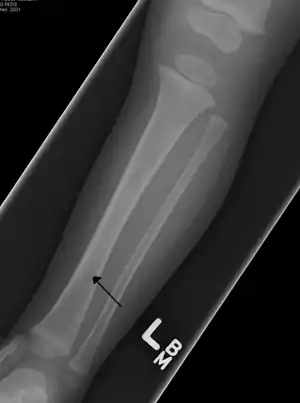
Toddler's fracture
Toddler's fractures are bone fractures of the distal (lower) part of the shin bone (tibia) in toddlers (aged 9 months-3 years) and other young children (less than 8 years).[1] The fracture is found in the distal two thirds of the tibia in 95% of cases,[1] is undisplaced and has a spiral pattern. It occurs after low-energy trauma, sometimes with a rotational component.
| Toddler's fracture | |
|---|---|
| Other names | Childhood accidental spiral tibial (CAST) fractures |
 | |
| A toddler's fracture | |
| Specialty | Orthopedic |
Pathophysiology
The proposed mechanism involves shear stress and lack of displacement due to the periosteum that is relatively strong compared to the elastic bone in young children.[2]
Diagnosis
Typical symptoms include pain, refusing to walk or bear weight and limping -bruising and deformity are absent. On clinical examination, there can be warmth and swelling over the fracture area, as well as pain on bending the foot upwards (dorsiflexion). The initial radiographical images may be inconspicuous (a faint oblique line) and often even completely normal.[3] After 1–2 weeks however, callus formation develops. The condition can be mistaken for osteomyelitis, transient synovitis or even child abuse. Contrary to CAST fractures, non-accidental injury typically affect the upper two-thirds or midshaft of the tibia.
Other possible fractures in this area, occurring in the cuboid, calcaneus, and fibula, can be associated or can be mistaken for a toddler's fracture.[4] In some cases, an internal oblique radiography and radionuclide imaging can add information to anterior-posterior and lateral views.[5][6] However, since treatment can also be initiated in the absence of abnormalities, this appears to have little value in most cases. It could be useful in special cases such as children with fever, those without a clear trauma or those in which the diagnosis remains unclear.[3][7] Recently, ultrasound has been suggested as a helpful diagnostic tool.[8]
Treatment
Treatment consist of a long leg orthopedic cast for several weeks.[3]
History
The condition was initially recognised by Dunbar and co-workers in 1964.[9] A new terminology has been proposed, which defines toddler's fracture as a subset of childhood accidental spiral tibial (CAST) fractures.[1]
References
- Mellick LB, Milker L, Egsieker E (October 1999). "Childhood accidental spiral tibial (CAST) fractures". Pediatr Emerg Care. 15 (5): 307–9. doi:10.1097/00006565-199910000-00001. PMID 10532655.
- Sarmah A (April 1995). "'Toddler's fracture'? A recognised entity". Arch. Dis. Child. 72 (4): 376. doi:10.1136/adc.72.4.376-a. PMC 1511261. PMID 7763080.
- Halsey MF, Finzel KC, Carrion WV, Haralabatos SS, Gruber MA, Meinhard BP (2001). "Toddler's fracture: presumptive diagnosis and treatment". J Pediatr Orthop. 21 (2): 152–6. doi:10.1097/00004694-200103000-00003. PMID 11242240.
- Donnelly LF (September 2000). "Toddler's fracture of the fibula". AJR Am J Roentgenol. 175 (3): 922. doi:10.2214/ajr.175.3.1750922. PMID 10954515.
- De Boeck K, Van Eldere S, De Vos P, Mortelmans L, Casteels-Van Daele M (January 1991). "Radionuclide bone imaging in toddler's fracture". Eur. J. Pediatr. 150 (3): 166–9. doi:10.1007/BF01963558. PMID 2044585. S2CID 11284378.
- John SD, Moorthy CS, Swischuk LE (1997). "Expanding the concept of the toddler's fracture". Radiographics. 17 (2): 367–76. doi:10.1148/radiographics.17.2.9084078. PMID 9084078.
- Miller JH, Sanderson RA (December 1988). "Scintigraphy of toddler's fracture". J. Nucl. Med. 29 (12): 2001–3. PMID 3193212.
- Lewis D, Logan P (May 2006). "Sonographic diagnosis of toddler's fracture in the emergency department". J Clin Ultrasound. 34 (4): 190–4. doi:10.1002/jcu.20192. PMID 16615049.
- Dunbar JS, Owen HF, Nogrady MB, McLeese R (September 1964). "Obscure tibial fracture of infants -- the toddler's fracture". J Can Assoc Radiol. 15: 136–44. PMID 14212071.