Chance fracture
A Chance fracture is a type of vertebral fracture that results from excessive flexion of the spine.[8] Symptoms may include abdominal bruising (seat belt sign), or less commonly paralysis of the legs.[4][9] In around half of cases there is an associated abdominal injury such as a splenic rupture, small bowel injury, pancreatic injury, or mesenteric tear.[3][5] Injury to the bowel may not be apparent in the first day.[10]
| Chance fracture | |
|---|---|
| Other names | Chance fracture of the spine,[1] flexion distraction fracture,[2] lap seat belt fracture[3] |
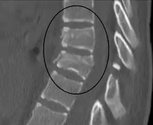 | |
| A Chance fracture of T10 and fracture of T9 due to a seatbelt during an MVC. | |
| Specialty | Emergency medicine |
| Symptoms | Abdominal bruising, paralysis of the legs[4] |
| Complications | Splenic rupture, small bowel injury, mesenteric tear[3][5] |
| Risk factors | Head-on motor vehicle collision in which a person is only wearing a lap belt[2] |
| Diagnostic method | Medical imaging (X-ray, CT scan)[1] |
| Differential diagnosis | Compression fracture, burst fracture[6] |
| Treatment | Bracing, surgery[1] |
| Frequency | Rare[7] |
The cause is classically a head-on motor vehicle collision in which the affected person is wearing only a lap belt.[2] Being hit in the abdomen with an object like a tree or a fall may also result in this fracture pattern.[11][9] It often involves disruption of all three columns of the vertebral body (anterior, middle, and posterior).[7][6] The most common area affected is the lower thoracic and upper lumbar spine.[6] A CT scan is recommended as part of the diagnostic work-up to detect any potential abdominal injuries.[5] The fracture is often unstable.[1]
Treatment may be conservative with the use of a brace or via surgery.[1] The fracture is currently rare.[7] It was first described by G. Q. Chance, a radiologist from Manchester, UK, in 1948.[3][12] The fracture was more common in the 1950s and 1960s before shoulder harnesses became common.[3][5]
Mechanism
In some Chance fractures there is a transverse break through the bony spinous process while in others there is a tear of the supraspinous ligament, ligamentum flavum, interspinous ligament, and posterior longitudinal ligament.[9]
Diagnosis
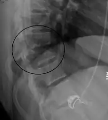
On plain X-ray, a Chance fracture may be suspected if two spinous processes are excessively far apart.[9]
A CT scan of the chest, abdomen, and pelvis is recommended as part of the diagnostic work-up to detect any potential abdominal injuries.[5][9] MRI may also be useful.[9] The fracture is often unstable.[1]
History
It was first described by G. Q. Chance, a radiologist from Manchester, UK, in 1948.[3][12] The fracture was more common in the 1950s and 1960s before shoulder harnesses became common.[3][5]
References
- "Wheeless' Textbook of Orthopaedics". Wheeless Online. Retrieved 29 May 2018.
- "Fractures of the Thoracic and Lumbar Spine". OrthoInfo - AAOS. Retrieved 29 May 2018.
- Yochum TR, Rowe LJ (2004). essentials of skeletal radiology. Lippincott Williams & Wilkins. p. 674.
- Eberhardt CS, Zand T, Ceroni D, Wildhaber BE, La Scala G (May 2016). "The Seatbelt Syndrome-Do We Have a Chance?: A Report of 3 Cases With Review of Literature". Pediatric Emergency Care. 32 (5): 318–22. doi:10.1097/PEC.0000000000000527. PMID 26087444. S2CID 25657579.
- Patel VV, Burger E, Brown CW (2010). Spine Trauma: Surgical Techniques. Springer Science & Business Media. p. 67. ISBN 9783642036941.
- Provenzale JM, Nelson RC, Vinson EN (2012). Duke Radiology Case Review: Imaging, Differential Diagnosis, and Discussion. Lippincott Williams & Wilkins. p. 247. ISBN 9781451180602.
- Marincek B, Dondelinger RF (2007). Emergency Radiology: Imaging and Intervention. Springer Science & Business Media. p. 152. ISBN 9783540689089.
- Masudi T, McMahon HC, Scott JL, Lockey AS (2017). "Seat belt-related injuries: A surgical perspective". Journal of Emergencies, Trauma, and Shock. 10 (2): 70–73. doi:10.4103/0974-2700.201590. PMC 5357874. PMID 28367011.
- Pope TL (2012). Harris & Harris' Radiology of Emergency Medicine. Lippincott Williams & Wilkins. p. 290. ISBN 9781451107203.
- Hopkins R, Peden C, Gandhi S (2009). Radiology for Anaesthesia and Intensive Care. Cambridge University Press. p. 114. ISBN 9781139482486.
- Hsu JD, Michael JW, Fisk JR, et al. (American Academy of Orthopaedic Surgeons) (2008). AAOS Atlas of Orthoses and Assistive Devices. Elsevier Health Sciences. p. 142. ISBN 978-0323039314.
- Chance GQ (September 1948). "Note on a type of flexion fracture of the spine". The British Journal of Radiology. 21 (249): 452–453. doi:10.1259/0007-1285-21-249-452. PMID 18878306.