Ankle fracture
An ankle fracture is a break of one or more ankle bones.[1] Symptoms may include pain, swelling, bruising, and an inability to walk on the leg.[1] Complications may include an associated high ankle sprain, compartment syndrome, decreased range of motion, and malunion.[1][2]
| Ankle fracture | |
|---|---|
| Other names | Broken ankle[1] |
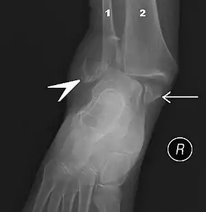 | |
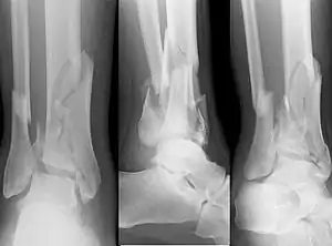
| Fracture of both sides of the ankle with dislocation as seen on anteroposterior X-ray. (1) fibula, (2) tibia, (arrow) medial malleolus, (arrowhead) lateral malleolus | |
| Specialty | Orthopedics |
| Symptoms | Pain, swelling, bruising, inability to walk[1] |
| Complications | High ankle sprain, compartment syndrome, decreased range of motion, malunion[1][2] |
| Usual onset | Young males, older females[2] |
| Types | Lateral malleolus, medial malleolus, posterior malleolus, bimalleolar, trimalleolar[1] |
| Causes | Rolling the ankle, blunt trauma[2] |
| Diagnostic method | X-rays based on the Ottawa ankle rule[2] |
| Differential diagnosis | Rheumatoid arthritis, gout, septic arthritis, Achilles tendon rupture[2] |
| Treatment | Splinting, casting, surgery[1] |
| Frequency | ~1 per 1000/year[2] |
The cause may include excessive stress on the joint such as from rolling an ankle or blunt trauma.[2][1] Types include lateral malleolus, medial malleolus, posterior malleolus, bimalleolar, and trimalleolar fractures.[1] The need for X-rays may be determined by the Ottawa ankle rule.[2]
Treatment is with splinting, casting, or surgery.[1] Ruling out other injuries may also be required.[2] Significant recovery generally occurs within four months; however, completely recovery may take up to two years.[1] They occur in about 1.7 per 1000 adults and 1 per 1000 children per year.[3][2] The occur most commonly in young males and older females.[2]
Signs and symptoms
Symptoms of an ankle fracture can be similar to those of ankle sprains (pain), though typically they are often more severe by comparison. It is exceedingly rare for the ankle joint to dislocate in the presence of ligamentous injury alone. However, in the setting of an ankle fracture the talus can become unstable and subluxate or dislocate. Patients may notice ecchymosis ("black and blue" coloraction from bleeding under the skin), or there may be an abnormal position, alignment, gross instability, or lack of normal motion secondary to pain. In a displaced fracture the skin is sometimes tented over a sharp edge of broken bone. The sharp fragments of broken bone sometimes tear the skin and form a laceration that communicates with the broken bone or joint space. This is known as an 'open' fracture and has a high incidence of infection if not promptly treated. Nearly all displaced ankle fractures are now treated surgically to insure proper alignment of the displaced fragments.
Diagnosis
On clinical examination, it is important to evaluate the exact location of the pain, the range of motion and the condition of the nerves and vessels. It is important to palpate the calf bone (fibula) because there may be an associated fracture proximally (Maisonneuve fracture), and to palpate the sole of the foot to look for a Jones fracture at the base of fifth metatarsal (avulsion fracture).
Evaluation of ankle injuries for fracture is done with the Ottawa ankle rules, a set of rules that were developed to minimize unnecessary X-rays. There are three x-ray views in a complete ankle series: anteroposterior, lateral, and oblique (or "mortise view"). The mortise view an anteroposterior x-ray taken with the ankle internally rotated until the lateral malleolus is on the same horizontal plane as the medial malleolus, and a line drawn through both malleoli would be parallel to the tabletop, resulting in a position where there normally is no superimposition of tibia and fibula on each other.[4] It usually requires 10 to 20 degrees of internal rotation.[4]
X-ray
On X-rays, there can be a fracture of the medial malleolus, the lateral malleolus, or of the anterior/posterior margin of the distal tibia. The posterior margin (known as the posterior malleolus) is much more frequently injured than the anterior aspect of the distal tibia. If both the lateral and medial malleoli are broken, this is called a bimalleolar fracture (some of them are called Pott's fractures). If the posterior malleolus is also fractured, this is called a trimalleolar fracture.
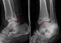 A triplane fracture of the ankle as seen on plain X-ray
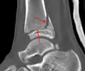 A triplane fracture of the ankle as seen on CT
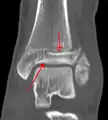 A triplane fracture of the ankle as seen on CT
Classification
There are several classification schemes for ankle fractures:
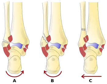
- The Lauge-Hansen classification categorises fractures based on the mechanism of the injury as it relates to the position of the foot and the deforming force (most common type is supination-external rotation)
- The Danis-Weber classification categorises ankle fractures by the level of the fracture of the distal fibula (type A = below the syndesmotic ligament, type B = at its level, type C = above the ligament), with use in assessing injury to the syndesmosis and the interosseous membrane
- The Herscovici classification categorises medial malleolus fractures of the distal tibia based on level.
- The Ruedi-Allgower classification categorizes pilon fractures of the distal tibia.
Fracture types
- Pilon fracture (Plafond fracture), a fracture of the distal part of the tibia, involving its articular surface at the ankle joint.
- Wagstaffe-Le Fort avulsion fracture¨, a vertical fracture of the antero-medial part of the distal fibula with avulsion of the anterior tibiofibular ligament.[5]
- Tillaux fracture, a Salter–Harris type III fracture through the anterolateral aspect of the distal tibial epiphysis.[6]
- Triplane fractures are a special type of fracture that involves the immature skeleton. It has a coronal plane in the metaphysis, an axial plane in the physis and a sagittal plane in the epiphysis.[7]

Treatment

Treatment of ankle fractures is dictated by the stability of the ankle joint. Certain fractures patterns are deemed stable, and may be treated similar to ankle sprains. All other types require surgery, most often an open reduction and internal fixation (ORIF), which is usually performed with permanently implanted metal hardware that holds the bones in place while the natural healing process occurs. A cast or splint will be required to immobilize the ankle following surgery.
In children, recovery may be faster with an ankle brace rather than a full cast in those with otherwise stable fractures.[3]
Epidemiology
In children ankle fractures occur in about 1 per 1000 per year.[3]
See also
References
- "Ankle Fractures (Broken Ankle) - OrthoInfo - AAOS". www.orthoinfo.org. Retrieved 20 June 2019.
- Wire, Jessica (9 May 2019). "Ankle Fractures". StatPearls. PMID 31194464.
- Yeung, DE; Jia, X; Miller, CA; Barker, SL (1 April 2016). "Interventions for treating ankle fractures in children". The Cochrane Database of Systematic Reviews. 4: CD010836. doi:10.1002/14651858.CD010836.pub2. PMC 7111433. PMID 27033333.
- Ankle Injuries: A Sprained Ankle? Radiology Cases in Pediatric Emergency Medicine Volume 3, Case 3 Alson S. Inaba MD Kapiolani Medical Center For Women And Children University of Hawaii John A. Burns School of Medicine. Retrieved May 2011
- Tim B Hunter; Leonard F Peltier; Pamela J Lund (2000). "Musculoskeletal Eponyms: Who Are Those Guys?" (PDF). RadioGraphics. 20: 829. doi:10.1148/radiographics.20.3.g00ma20819. PMID 10835130. Retrieved 2009-11-13.
- "Wheeless Online". Retrieved 30 October 2014.
- Hirsch M, et al. Understanding triplane distal tibia fractures. http://dx.doi.org/10.1016/j.rchira.2016.09.002
External links
| Classification |
|---|
| Wikimedia Commons has media related to Fractures of the human ankles. |
| Wikiquote has quotations related to: Ankle fracture |