Bronchial artery
In human anatomy, the bronchial arteries supply the lungs with nutrition and oxygenated blood. Although there is much variation, there are usually two bronchial arteries that run to the left lung, and one to the right lung.
| Bronchial artery | |
|---|---|
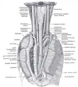 Bronchial artery labeled at center right | |
| Details | |
| Source | Thoracic aorta |
| Vein | Bronchial veins |
| Supplies | Lungs |
| Identifiers | |
| Latin | Arteriae bronchiales, rami bronchiales partis thoracicae aortae |
| MeSH | D001981 |
| TA98 | A12.2.11.002 |
| TA2 | 4579 |
| FMA | 68109 71536, 68109 |
| Anatomical terminology | |
Structure
The left bronchial arteries (superior and inferior) usually arise directly from the thoracic aorta.[1]
The single right bronchial artery usually arises from one of the following:
- 1) the thoracic aorta at a common trunk with the right 3rd posterior intercostal artery
- 2) the superior bronchial artery on the left side
- 3) any number of the right intercostal arteries mostly the third right posterior.
Function
The bronchial arteries supply blood to the bronchi and connective tissue of the lungs. They travel with and branch with the bronchi, ending about at the level of the respiratory bronchioles. They anastomose with the branches of the pulmonary arteries, and together, they supply the visceral pleura of the lung in the process.
Note that much of the oxygenated blood supplied by the bronchial arteries is returned via the pulmonary veins rather than the bronchial veins. As a consequence, blood returning to the left heart is slightly less oxygenated than blood found at the level of the pulmonary capillary beds.
Each bronchial artery also has a branch that supplies the esophagus.
Comparison with pulmonary arteries
It is easy to confuse the bronchial arteries with the pulmonary arteries, because they both supply the lungs with blood, but there are important differences:
| artery | function | circulation | diameter |
| pulmonary arteries | supplies deoxygenated blood pumped from the right ventricle | pulmonary circulation | relatively large |
| bronchial arteries | supplies oxygenated blood pumped from the left ventricle | systemic circulation | relatively small |
Clinical significance
The bronchial arteries are typically enlarged and tortuous in chronic pulmonary thromboembolic hypertension.[2]
With modern surgical techniques, bronchial anastomoses heal well without bronchial artery reconnection. Largely for this reason, bronchial artery circulation is usually sacrificed during lung transplants, instead relying on the persistence of a microcirculation (presumably arising from the deoxygenated pulmonary circulation) to provide perfusion to the airways.[3]
Aneurysms of the bronchial artery may mimic aortic aneurysms.[4] Bronchial artery embolisation (BAE) is catheter insertion into a bronchial artery to treat hemopytsis (coughing blood).[5][6]
The bronchial arteries and their supply of nutrients to the lungs are also attributed to the observation that an occluded (either ligated or by an embolus) pulmonal artery very rarely results in lung infarction.[7] The bronchial arteries can maintain a supply of oxygenated blood to lung tissue.
References
- Leslie, Kevin O.; Wick, Mark R. (2018-01-01), Leslie, Kevin O.; Wick, Mark R. (eds.), "1 - Lung Anatomy", Practical Pulmonary Pathology: A Diagnostic Approach (Third Edition), Elsevier, pp. 1–14.e2, doi:10.1016/b978-0-323-44284-8.00001-6, ISBN 978-0-323-44284-8, retrieved 2020-11-17
- Kauczor H, Schwickert H, Mayer E, Schweden F, Schild H, Thelen M (1994). "Spiral CT of bronchial arteries in chronic thromboembolism". J Comput Assist Tomogr. 18 (6): 855–61. doi:10.1097/00004728-199411000-00002. PMID 7962789.
- Nowak et al. Bronchial Artery Revascularization affects Graft Recovery after Lung Transplant. AJRCCM Vol 165. Number 2, Jan 2002
- Vernhet H, Bousquet C, Jean B, et al. (1999). "Bronchial aneurysms mimicking aortic aneurysms: endovascular treatment in two patients". Cardiovasc Intervent Radiol. 22 (3): 254–7. doi:10.1007/s002709900378. PMID 10382061.
- Ennis James, W. (2019-01-01), Baughman, Robert Phillip; Valeyre, Dominique (eds.), "Chapter 23 - Nonpharmacological Therapy for Pulmonary Sarcoidosis", Sarcoidosis, Philadelphia: Elsevier, pp. 277–284, doi:10.1016/b978-0-323-54429-0.00023-9, ISBN 978-0-323-54429-0, retrieved 2020-11-17
- Garwood, Susan; Strange, Charlie; Sahn, Steven A. (2008-01-01), Parrillo, Joseph E.; Dellinger, R. Phillip (eds.), "Chapter 47 - Massive Hemoptysis", Critical Care Medicine (Third Edition), Philadelphia: Mosby, pp. 929–948, doi:10.1016/b978-032304841-5.50049-2, ISBN 978-0-323-04841-5, retrieved 2020-11-17
- Fishman, Alfred P. (1961). "The Clinical Significance of the Pulmonary Collateral Circulation". Circulation. American Heart Association. 24: 677–690. doi:10.1161/01.CIR.24.3.677. eISSN 1524-4539. ISSN 0009-7322.
External links
- Anatomy figure: 21:06-06 at Human Anatomy Online, SUNY Downstate Medical Center - "Branches of the ascending aorta, arch of the aorta, and the descending aorta."
- Histology image: 13903loa – Histology Learning System at Boston University
- Bronchial arteries - anatquest.nlm.nih.gov.
Vital volume is 4600ml
Residual volume is 1200ml
| Authority control |
|---|