Superior epigastric artery
In human anatomy, the superior epigastric artery is a blood vessel that carries oxygenated blood to the abdominal wall,[1][2] and upper rectus abdominis muscle.[1]
| Superior epigastric artery | |
|---|---|
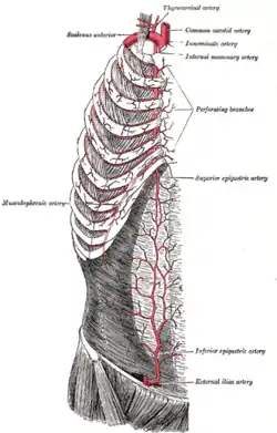
 Superior epigastric artery, internal thoracic artery and inferior epigastric artery. (Superior epigastric artery is labeled at right center.) | |
| Details | |
| Source | internal thoracic |
| Vein | superior epigastric vein |
| Identifiers | |
| Latin | arteria epigastrica superior |
| TA98 | A12.2.08.041 |
| TA2 | 4588 |
| FMA | 10646 |
| Anatomical terminology | |
Structure
The superior epigastric artery arises from the internal thoracic artery (referred to as the internal mammary artery in the accompanying diagram).[3][4] It anastomoses with the inferior epigastric artery at the umbilicus.[3] Along its course, it is accompanied by a similarly named vein, the superior epigastric vein.
Function
Where it anastomoses, the superior epigastric artery supplies the anterior part of the abdominal wall,[1][2] upper rectus abdominis muscle,[1] and some of the diaphragm.
Collateralization in disease
Vascular disease
The superior epigastric arteries, inferior epigastric arteries, internal thoracic arteries and left subclavian artery and right subclavian artery / brachiocephalic are collateral vessels to the thoracic aorta and abdominal aorta. If the abdominal aorta develops a significant stenosis and/or blockage (as may be caused by atherosclerosis), this collateral pathway may develop sufficiently, over time, to supply blood to the lower limbs.[5]
Coarctation of the aorta
A congenitally narrowed aorta, due to coarctation, is often associated with a significant enlargement of the internal thoracic and epigastric arteries.[6]
References
- Shell, Dan H.; Vásconez, Luis O.; de la Torre, Jorge I.; Chin, Gloria; Weinzweig, Norman (January 1, 2010), Weinzweig, Jeffrey (ed.), "Chapter 91 - Abdominal Wall Reconstruction", Plastic Surgery Secrets Plus (Second Edition), Philadelphia: Mosby, pp. 594–604, doi:10.1016/b978-0-323-03470-8.00091-0, ISBN 978-0-323-03470-8, retrieved November 22, 2020
- DiEdwardo, Christine A.; Caterson, Stephanie A.; Barrall, David T. (January 1, 2010), Weinzweig, Jeffrey (ed.), "Chapter 80 - Abdominoplasty", Plastic Surgery Secrets Plus (Second Edition), Philadelphia: Mosby, pp. 532–537, doi:10.1016/b978-0-323-03470-8.00080-6, ISBN 978-0-323-03470-8, retrieved November 22, 2020
- Castro Ferreira, Marcus; Henrique Ishida, Luis; Munhoz, Alexandre (January 1, 2009), Wei, Fu-Chan; Mardini, Samir (eds.), "CHAPTER 19 - Rectus flap", Flaps and Reconstructive Surgery, Edinburgh: W.B. Saunders, pp. 207–223, doi:10.1016/b978-0-7216-0519-7.00019-8, ISBN 978-0-7216-0519-7, retrieved November 22, 2020
- Ahmed, Abdul (January 1, 2017), Brennan, Peter A.; Schliephake, Henning; Ghali, G. E.; Cascarini, Luke (eds.), "37 - Common Free Vascularized Flaps: The Rectus Abdominis", Maxillofacial Surgery (Third Edition), Churchill Livingstone, pp. 533–542, doi:10.1016/b978-0-7020-6056-4.00038-1, ISBN 978-0-7020-6056-4, retrieved November 22, 2020
- Yurdakul M, Tola M, Ozdemir E, Bayazit M, Cumhur T (April 2006). "Internal thoracic artery-inferior epigastric artery as a collateral pathway in aortoiliac occlusive disease". J. Vasc. Surg. 43 (4): 707–13. doi:10.1016/j.jvs.2005.12.042. PMID 16616225.
- Huhmann W, Kunitsch G, Dalichau H (1976). "[Coarctation of the aorta on the plain chest x-ray (author's transl)]". Dtsch Med Wochenschr. 101 (41): 1477–81. doi:10.1055/s-0028-1104294. PMID 964150.
External links
- Anatomy photo:18:07-0103 at the SUNY Downstate Medical Center - "Thoracic wall: Branches of the Internal Thoracic Artery"
- Anatomy figure: 35:04-03 at Human Anatomy Online, SUNY Downstate Medical Center - "Incisions and the contents of the rectus sheath. "