Clivus (anatomy)
The clivus (Latin for "slope") is a bony[1] part of the cranium at the skull base, a shallow depression behind the dorsum sellæ that slopes obliquely backward. It forms a gradual sloping process at the anterior most portion of the basilar occipital bone at its junction with the sphenoid bone. On axial planes, it sits just posterior to the sphenoid sinuses. Just lateral to the clivus bilaterally is the foramen lacerum (the internal carotid artery reaches the middle cranial fossa above the foramen lacerum), proximal to its anastomosis with the Circle of Willis. Posterior to the clivus is the basilar artery.
| Clivus | |
|---|---|
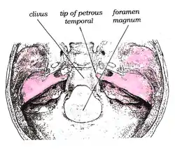 Superior view of the clivus | |
| Details | |
| Identifiers | |
| Latin | Clivus |
| TA98 | A02.1.00.051 A02.1.04.006 |
| TA2 | 454 |
| FMA | 54376 |
| Anatomical terms of bone | |
The pons sits on the clivus.
Clivus is also used as an abbreviated term for the clivus ocularis which is the sloping inner wall of the retina as it dips into the foveola in the macula of the eye. For this reason, and to disambiguate, the clivus is sometimes referred to as the Blumenbach clivus.
Clinical importance
The abducens nerve (cranial nerve VI) tracks along the clivus during its course. Increased intracranial pressure can trap the nerve at this point and cause signs of palsy.
Clivus is also the site for chordoma (a rare malignant tumour.)
Surgery for lesions involving the clivus and surrounding structures have traditionally been approached via extended subfrontal transbasal, anterior transfacial, lateral transtemporal, far-lateral approaches, and staged approaches.[2] These approaches are limited in that they often require extensive bone removal and brain retraction while placing critical neurovascular structures between the surgeon and the site of pathology. It has been proposed that these limitations are mitigated by significant advancements in the use of endoscopic endonasal surgery. Contemporary surgical approaches involving extended endoscopic endonasal approaches to the clivus have been increasingly described by several groups, and have been shown to be a safe and effective strategy for the surgical management of a variety of benign and malignant lesions.[2]
Relation of the clivus and dens
The clivus is an important landmark for checking for anatomical atlanto-occipital alignment; the clivus, when viewed on a lateral C-spine X-ray, forms a line which, if extended, is known as Wackenheim's clivus line. Wackenheim's clivus line should pass through the dens of the axis or be tangential to it.[3]
Additional Images
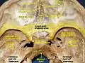 Clivus
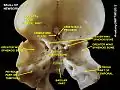 Clivus
See also
References
This article incorporates text in the public domain from page 148 of the 20th edition of Gray's Anatomy (1918)
- Drake, Richard L. Gray's Anatomy for Students 3rd Ed. p. 868.
- Little, Ryan E.; Taylor, Robert J.; Miller, Justin D.; Ambrose, Emily C.; Germanwala, Anand V.; Sasaki-Adams, Deanna M.; Ewend, Matthew G.; Zanation, Adam M. (August 2014). "Endoscopic endonasal transclival approaches: case series and outcomes for different clival regions". Journal of Neurological Surgery. Part B, Skull Base. 75 (4): 247–254. doi:10.1055/s-0034-1371522. ISSN 2193-6331. PMC 4108492. PMID 25093148.
- McKenna DA, Roche CJ, Lee KW, Torreggiani WC, Duddalwar VA. Atlanto-occipital dislocation: case report and discussion. Can J Emerg Med 2006; 8(1):50-3. Available at: link Archived 2007-09-27 at the Wayback Machine and link Archived 2007-09-27 at the Wayback Machine. Accessed on: December 7, 2006.
External links
- Anatomy photo:22:os-0913 at the SUNY Downstate Medical Center - "Osteology of the Skull: Internal Surface of Skull"
- Diagram at uwo.ca
| Authority control |
|---|