Glycerate dehydrogenase
In enzymology, a glycerate dehydrogenase (EC 1.1.1.29) is an enzyme that catalyzes the chemical reaction
- (D)-glycerate + NAD+ hydroxypyruvate + NADH + H+
| Glycerate dehydrogenase | |||||||||
|---|---|---|---|---|---|---|---|---|---|
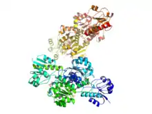
 Glyoxylate reductase/Hydroxypyruvate reductase. Quaternary structure of 2 homodimers of GRHPR bound to NADPH and (D)-glycerate. | |||||||||
| Identifiers | |||||||||
| EC number | 1.1.1.29 | ||||||||
| CAS number | 9028-37-9 | ||||||||
| Databases | |||||||||
| IntEnz | IntEnz view | ||||||||
| BRENDA | BRENDA entry | ||||||||
| ExPASy | NiceZyme view | ||||||||
| KEGG | KEGG entry | ||||||||
| MetaCyc | metabolic pathway | ||||||||
| PRIAM | profile | ||||||||
| PDB structures | RCSB PDB PDBe PDBsum | ||||||||
| Gene Ontology | AmiGO / QuickGO | ||||||||
| |||||||||
Thus, the two substrates of this enzyme are (R)-glycerate and NAD+, whereas its 3 products are hydroxypyruvate, NADH, and H+. However, in nature these enzymes have the ability to catalyze the reverse reaction as well. That is, hydroxypyruvate, NADH, and H+ can act as the substrates while (R)-glycerate and NAD+ are formed as products. Additionally, NADPH can take the place of NADH in this reaction.[1]
This enzyme belongs to the family of oxidoreductases, specifically those acting on the CH-OH group of donor with NAD+ or NADP+ as acceptor. The systematic name of this enzyme class is (R)-glycerate:NAD+ oxidoreductase. Other names in common use include D-glycerate dehydrogenase, and hydroxypyruvate reductase (due to the reversibility of the reaction). This enzyme participates in glycine, serine and threonine metabolism and glyoxylate and dicarboxylate metabolism.
Enzyme structure
This class of enzyme is part of a larger superfamily of enzymes known as D-2-hydroxy-acid dehydrogenases.[2] Many organisms from Hyphomicrobium methylovorum to humans have some form of the glycerate dehydrogenase protein. There are currently several structures that have been solved for this class of enzyme including those for the two mentioned above with PDB access code 1GDH, D-glycerate dehydrogenase, and the human homolog Glyoxylate reductase/Hydroxypyruvate reductase (GRHPR), 2WWR.
These studies have yielded a better understanding of the structure and function of these enzymes. It has been shown that these proteins are homodimeric enzymes.[3] This means that 2 identical proteins are linked forming one larger complex. The active site is found in each subunit between the two distinct α/β/α globular domains, the substrate binding domain and the coenzyme binding domain.[2] This coenzyme binding domain is slightly larger than the substrate binding domain and contains a NAD(P) Rossmann fold along with the "dimerisation loop" which holds the two subunits of the homodimer together.[2] In addition to linking the two proteins together, the "dimerisation loop" of each subunit protrudes into the active site of the other subunit increasing the specificity of the enzyme, by preventing the binding of pyruvate as a substrate. Hydroxypyruvate is still able to bind to the active site due to extra stabilization from hydrogen bonds with neighboring amino-acid residues.[2]
Glyoxylate reductase/Hydroxypyruvate reductase
Biological relevance
Glyoxylate reductase/Hydroxypyruvate reductase (GRHPR) is the glycerate dehydrogenase found, predominantly in the liver, of humans encoded by the gene GRHPR.[4] Under physiological conditions, the production of D-glycerate is favored over its consumption as a substrate. It can then be converted to 2-phosphoglycerate,[5] which can then enter into glycolysis, gluconeogenesis, or the serine pathway.[6][7]
As the name suggests, in addition to the glycerate dehydrogenase and hydroxypyruvate reductase activity, the protein also exhibits glyoxylate reductase activity.[2] The ability of GRHPR to reduce glyoxylate to glycolate is found in other glycerate dehydrogenase homologs as well.[1] This is important for the intracellular regulation of glyoxylate levels, which has important medical ramifications. As mentioned earlier, these enzymes have the ability to use either NADH or NADPH as the coenzyme. This gives them an advantage over other enzymes that can only use a single form of the coenzyme. Lactate dehydrogenase(LDH) is one such enzyme that directly competes with GRHPR for substrates and converts glyoxylate to oxalate. However, due to the relatively large concentration of NADPH compared to NADH under normal cellular concentration, the GRHPR activity is greater than that of LDH so the production of glycolate is dominant.[2]
Medical relevance
Primary hyperoxaluria is a condition that results in the overproduction of oxalate which combines with calcium to generate calcium oxalate, the main component of kidney stones.[8][9] Primary Hyperoxaluria type 2 is caused by any one of several mutations to the GRHPR gene and results in the accumulation of calcium oxalate in the kidneys, bones, and many other organs.[8] The mutations to GRHPR prevent it from converting glyoxylate to glycolate, leading to a build-up of glyoxylate. This excess glyoxylate is then oxidized by lactate dehydrogenase to produce the oxalate that is characteristic of hyperoxaluria.
References
- Schaftingen, Emile; Fancois Van Hoof (Feb 1989). "Coenzyme Specificity of Mammalian liver D-glycerate dehydrogenase". European Journal of Biochemistry. 186 (1–2): 355–359. doi:10.1111/j.1432-1033.1989.tb15216.x. PMID 2689175.
- Booth, Michael; R. Connors; G. Rumsby; R. Leo Brady (18 May 2006). "Structural basis of substrate specificity in human glyoxylate reductase/hydroxypyruvate reductase". Journal of Molecular Biology. 360 (1): 178–189. doi:10.1016/j.jmb.2006.05.018. PMID 16756993.
- Goldberg, JD; Yoshida, T; Brick, P (March 4, 1994). "Crystal structure of a NAD-dependent D-glycerate dehydrogenase at 2.4 Å resolution". Journal of Molecular Biology. 236 (4): 1123–40. doi:10.1016/0022-2836(94)90016-7. PMID 8120891.
- Cregeen, DP; Williams, EL; Hulton, S; Rumsby, G (December 2003). "Molecular analysis of the glyoxylate reductase (GRHPR) gene and description of mutations underlying primary hyperoxaluria type 2". Human Mutation. 22 (6): 497. doi:10.1002/humu.9200. PMID 14635115. S2CID 39645821.
- Liu, B; Hong, Y; Wu, L; Li, Z; Ni, J; Sheng, D; Shen, Y (September 2007). "A unique highly thermostable 2-phosphoglycerate forming glycerate kinase from the hyperthermophilic archaeon Pyrococcus horikoshii: gene cloning, expression and characterization". Extremophiles : Life Under Extreme Conditions. 11 (5): 733–9. doi:10.1007/s00792-007-0079-9. PMID 17563835. S2CID 38801171.
- Quayle, JR (February 1980). "Microbial assimilation of C1 compounds. The Thirteenth CIBA Medal Lecture". Biochemical Society Transactions. 8 (1): 1–10. doi:10.1042/bst0080001. PMID 6768606.
- O'Connor, ML; Hanson, RS (November 1975). "Serine transhydroxymethylase isoenzymes from a facultative methylotroph". Journal of Bacteriology. 124 (2): 985–96. doi:10.1128/JB.124.2.985-996.1975. PMC 235989. PMID 241747.
- Mitsimponas, KT; Wehrhan, T; Falk, S; Wehrhan, F; Neukam, FW; Schlegel, KA (December 2012). "Oral findings associated with primary hyperoxaluria type I". Journal of Cranio-Maxillo-Facial Surgery. 40 (8): e301-6. doi:10.1016/j.jcms.2012.01.009. PMID 22417769.
- "Primary hyperoxaluria". Retrieved 4 March 2013.
- Holzer H, Holldorf A (1957). "[Isolation of D-glycerate dehydrogenase, some properties of the enzyme and its application to the enzymic-optic determination of hydroxypyruvate in presence of pyruvate]". Biochem. Z. (in German). 329 (4): 292–312. PMID 13522707.
- Stafford HA, Magaldi A, Vennesland B (1954). "The enzymatic reduction of hydroxypyruvic acid to D-glyceric acid in higher plants". J. Biol. Chem. 207 (2): 621–9. PMID 13163046.
- Rumsby, G; Pagon, RA; Bird, TD; Dolan, CR; Stephens, K; Adam, MP (1993). "Primary Hyperoxaluria Type 2". PMID 20301742. Cite journal requires
|journal=(help) - Cramer, SD; Ferree, PM; Lin, K; Milliner, DS; Holmes, RP (October 1999). "The gene encoding hydroxypyruvate reductase (GRHPR) is mutated in patients with primary hyperoxaluria type II". Human Molecular Genetics. 8 (11): 2063–9. doi:10.1093/hmg/8.11.2063. PMID 10484776.