Major histocompatibility complex, class I-related
Major histocompatibility complex class I-related gene protein (MR1) is a non-classical MHC class I protein, that binds vitamine metabolites (intermediates of riboflavin synthesis) produced in certain types of bacteria. MR1 interacts with mucosal associated invariant T cells (MAIT).[5][6]
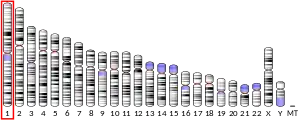


Gene location
MR1 is a protein that in humans is encoded by the MR1 gene and located on chromosome 1. Non-classical MHC class I genes are very often located on the same chromosome (mice chromosome 6, human chromosome 17) and interspaced within the same loci as the classical MHC genes. MR1 is located on another chromosome, the detailed gene analysis revealed that MR1 is a paralog originated by duplication of MHC locus on chromosome 6 (mice). This functional gene has been found in almost all mammals, proving the importance of MR1 across the mammalian kingdom and the fact that the duplication occurred early in the evolution of vertebrates.[5][7][8]
Another non-classical MHC class I CD1 is missing in certain species. There is 90% protein homology of the MR1 binding site between mice and humans. MR1 shares greater homology with classical MHC I class than with non-classical MHC class I. The human MR1 protein has 341 amino acid residues with a molecular weight of 39 366 Daltons.[9][5]
Structure
MR1, like other MHC class I molecules, is composed of α1, α2 and α3 domains. α1 and α2 interact and together bind the antigen. The ligand binding pocket is small and contains aromatic and basic residues. Its small size limits it to only binding molecules of a similarly small size. α3 interacts with β2 microglobulin.[5]
Many different isoforms of MR1 have been identified. Many of the identified proteins have a premature terminating codon which generates non-functional proteins. MR1B isoform lacks the α3 domain. The α3 domain interacts with β2 microglobulin. This interaction and binding of the antigen stabilize the MHC I molecule. In the case of MR1B β2 microglobulin is not needed for stabilization of the structure. MR1B is expressed on the cell surface. This isoform binds antigen via α1 and α2 interaction. Some bacteria are able to target specific β2 microglobulin that enable MHC I presentation. This might be a mechanism used to avoid bacterial immune evasion during bacterial infections.[5]
Antigen presentation
The MR1 protein is capable of binding to molecules derived from bacterial riboflavin biosynthesis, and then present them to MAIT for activation.[7][8]
MR1 is almost undetectable under physiological conditions, surface expression increase in cells infected by microbes. Due to the antigen necessity for MR1 stabilization. MR1 binds the intermediates of riboflavine synthesis. Human body can´t synthesize most of the vitamins, thus the presence of intermediates of riboflavin synthesis is a marker of non-self. Many bacteria are capable of vitamine synthesis.[10] The first discovered MR1 ligand was 6-formyl pterin (6-FP).[5]
Within cells, MR1 is mostly stored inside the endoplasmic reticulum (ER), where ligand binding occurs.[11] Inside the ER, MR1 is stabilised in a ligand-receptive conformation by chaperone proteins tapasin and TAP-binding protein related (TAPBPR) to facilitate ligand binding.[11] After antigen binding MR1 undergoes conformational change, associate with β2 microglobulin and is directed to the cell membrane.[12][5][10]
MR1 stimulation is needed for MAIT development in thymus, the mechanism of antigen presentation in thymus is not clear.[5]
MR1 ligands
The MR1 pocket is composed primary of aromatic and basic amino acids and the volume is small, thus suitable for binding small molecule ligands.
The ligands 5-OP-RU and 5-OE-RU are compounds derived from riboflavin biosynthesis that bind MR1 for presentation to MAIT cells for activation.[13] They are chemically unstable, but have been synthesised as chemical tools for studying MR1 biology.[14] Ac-6-FP (acetyl-6-formylpterin) and 6-FP (6-formylpterin) also bind MR1, but they do not activate MAIT cells.[8] There is an evidence, that MR1 can bind other antigens. MR1 was able to stimulate T lymphocytes in the presence of Streptoccocus pyogenes, that is unable to synthesise riboflavin. MR1 is important in the immune fight against cancer, because MR1 T lymphocytes were able to selectively kill various cancer cells.[15]
Clinical signficance
A study showed that MR1 T lymphocytes were able to kill various cancer cells in vivo and in vitro and ignored noncancerous cells. The MR1 recognition was independent of bacterial load.[16]
MR1 deficient mice had impaired early phase mucosal cytokine production and delayed recruitment of activated T lymphocytes into infection site.[10]
There has not been any evidence of self antigens stimulating MR1 autoreactive T cells in any autoimmune disease.[10]
References
- GRCh38: Ensembl release 89: ENSG00000153029 - Ensembl, May 2017
- GRCm38: Ensembl release 89: ENSMUSG00000026471 - Ensembl, May 2017
- "Human PubMed Reference:". National Center for Biotechnology Information, U.S. National Library of Medicine.
- "Mouse PubMed Reference:". National Center for Biotechnology Information, U.S. National Library of Medicine.
- Krovi SH, Gapin L (August 2016). "Structure and function of the non-classical major histocompatibility complex molecule MR1". Immunogenetics. 68 (8): 549–59. doi:10.1007/s00251-016-0939-5. PMC 5091073. PMID 27448212.
- McWilliam HE, Villadangos JA (September 2017). "How MR1 Presents a Pathogen Metabolic Signature to Mucosal-Associated Invariant T (MAIT) Cells". Trends in Immunology. 38 (9): 679–689. doi:10.1016/j.it.2017.06.005. PMID 28688841.
- Corbett AJ, Eckle SB, Birkinshaw RW, Liu L, Patel O, Mahony J, et al. (May 2014). "T-cell activation by transitory neo-antigens derived from distinct microbial pathways". Nature. 509 (7500): 361–5. Bibcode:2014Natur.509..361C. doi:10.1038/nature13160. PMID 24695216.
- Kjer-Nielsen L, Patel O, Corbett AJ, Le Nours J, Meehan B, Liu L, et al. (November 2012). "MR1 presents microbial vitamin B metabolites to MAIT cells" (PDF). Nature. 491 (7426): 717–23. Bibcode:2012Natur.491..717K. doi:10.1038/nature11605. PMID 23051753.
- "UniProt, Q95460".
- Mori L, Lepore M, De Libero G (May 2016). "The Immunology of CD1- and MR1-Restricted T Cells". Annual Review of Immunology. 34 (1): 479–510. doi:10.1146/annurev-immunol-032414-112008. PMID 26927205.
- McWilliam, Hamish E. G.; Mak, Jeffrey Y. W.; Awad, Wael; Zorkau, Matthew; Cruz-Gomez, Sebastian; Lim, Hui Jing; Yan, Yuting; Wormald, Sam; Dagley, Laura F.; Eckle, Sidonia B. G.; Corbett, Alexandra J. (2020-09-18). "Endoplasmic reticulum chaperones stabilize ligand-receptive MR1 molecules for efficient presentation of metabolite antigens". Proceedings of the National Academy of Sciences. doi:10.1073/pnas.2011260117. ISSN 0027-8424. PMID 32958637.
- McWilliam HE, Eckle SB, Theodossis A, Liu L, Chen Z, Wubben JM, et al. (May 2016). "The intracellular pathway for the presentation of vitamin B-related antigens by the antigen-presenting molecule MR1" (PDF). Nature Immunology. 17 (5): 531–7. doi:10.1038/ni.3416. PMID 27043408.
- Corbett AJ, Eckle SB, Birkinshaw RW, Liu L, Patel O, Mahony J, et al. (May 2014). "T-cell activation by transitory neo-antigens derived from distinct microbial pathways". Nature. 509 (7500): 361–5. doi:10.1038/nature13160. PMID 24695216.
- Mak JY, Xu W, Reid RC, Corbett AJ, Meehan BS, Wang H, et al. (March 2017). "Stabilizing short-lived Schiff base derivatives of 5-aminouracils that activate mucosal-associated invariant T cells". Nature Communications. 8 (1): 14599. doi:10.1038/ncomms14599. PMC 5344979. PMID 28272391.
- Karamooz E, Harriff MJ, Lewinsohn DM (December 2018). "MR1-dependent antigen presentation". Seminars in Cell & Developmental Biology. 84: 58–64. doi:10.1016/j.semcdb.2017.11.028. PMC 7061520. PMID 30449535.
- Crowther MD, Dolton G, Legut M, Caillaud ME, Lloyd A, Attaf M, et al. (February 2020). "Genome-wide CRISPR-Cas9 screening reveals ubiquitous T cell cancer targeting via the monomorphic MHC class I-related protein MR1". Nature Immunology. 21 (2): 178–185. doi:10.1038/s41590-019-0578-8. PMC 6983325. PMID 31959982.
Further reading
- Maruyama K, Sugano S (January 1994). "Oligo-capping: a simple method to replace the cap structure of eukaryotic mRNAs with oligoribonucleotides". Gene. 138 (1–2): 171–4. doi:10.1016/0378-1119(94)90802-8. PMID 8125298.
- Yamaguchi H, Hirai M, Kurosawa Y, Hashimoto K (September 1997). "A highly conserved major histocompatibility complex class I-related gene in mammals". Biochemical and Biophysical Research Communications. 238 (3): 697–702. doi:10.1006/bbrc.1997.7379. PMID 9325151.
- Suzuki Y, Yoshitomo-Nakagawa K, Maruyama K, Suyama A, Sugano S (October 1997). "Construction and characterization of a full length-enriched and a 5'-end-enriched cDNA library". Gene. 200 (1–2): 149–56. doi:10.1016/S0378-1119(97)00411-3. PMID 9373149.
- Riegert P, Wanner V, Bahram S (October 1998). "Genomics, isoforms, expression, and phylogeny of the MHC class I-related MR1 gene". Journal of Immunology. 161 (8): 4066–77. PMID 9780177.
- Parra-Cuadrado JF, Navarro P, Mirones I, Setién F, Oteo M, Martínez-Naves E (August 2000). "A study on the polymorphism of human MHC class I-related MR1 gene and identification of an MR1-like pseudogene". Tissue Antigens. 56 (2): 170–2. doi:10.1034/j.1399-0039.2000.560211.x. PMID 11019920.
- Miley MJ, Truscott SM, Yu YY, Gilfillan S, Fremont DH, Hansen TH, Lybarger L (June 2003). "Biochemical features of the MHC-related protein 1 consistent with an immunological function". Journal of Immunology. 170 (12): 6090–8. doi:10.4049/jimmunol.170.12.6090. PMID 12794138.
- Hempelmann A, Kumar S, Muralitharan S, Sander T (July 2006). "Myofibrillogenesis regulator 1 gene (MR-1) mutation in an Omani family with paroxysmal nonkinesigenic dyskinesia". Neuroscience Letters. 402 (1–2): 118–20. doi:10.1016/j.neulet.2006.03.048. PMID 16632198.