Olfactory glands
Olfactory glands, also known as Bowman's glands, are a type of nasal gland situated in the olfactory mucosa, beneath the olfactory epithelium, in the lamina propria, a connective tissue also containing fibroblasts, blood vessels and bundles of fine axons from the olfactory neurons.[1]
| Olfactory glands | |
|---|---|
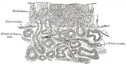 Section of the olfactory mucous membrane | |
| Details | |
| System | Olfactory system |
| Identifiers | |
| Latin | glandulae olfactoriae |
| TA98 | A15.1.00.003 |
| TA2 | 6733 |
| TH | H3.05.00.0.00026 |
| FMA | 77659 |
| Anatomical terminology | |
An olfactory gland consists of an acinus in the lamina propria and a secretory duct going out through the olfactory epithelium.
Electron microscopy studies show that olfactory glands contain cells with large secretory vesicles.[2] Olfactory glands secrete the gel-forming mucin protein MUC5AC.[3] They might secrete proteins such as lactoferrin, lysozyme, amylase and IgA, similarly to serous glands. The exact composition of the secretions from olfactory glands is unclear, but there is evidence that they produce odorant-binding protein.[4][5]
Function
The Olfactory glands are theses tubuloalveolar glands surrounded by the Olfactory receptor and the Sustentacular cell in the olfactory epithelium. These bulbus glands function to produce mucus to keep the olfactory epithelium moist as well as dissolve odor containing gases. A number of olfactory binding proteins are produced from the olfactory glands that help facilitate the transportation of odorants to the olfactory receptors. These cells exhibit the mRNA to transform growth factor α, stimulating the production of new olfactory receptor cells.
Mucus production from the olfactory glands also play an important role in protecting the surface of the epithelium from drying out.
Role in Olfactory System
Once air is inhaled through the nose and enters the nasal cavity, the odorant molecules travel all the way to the olfactory epithelium surface covered in olfactory cilia. These olfactory cilia are protected by a layer of mucus, produced by these olfactory glands, which trap these odorants, allowing them to make there way to the cilia and up to the olfactory receptors.
See also
References
This article incorporates text in the public domain from page 996 of the 20th edition of Gray's Anatomy (1918)
- Moran, David T.; Rowley Jc, 3rd; Jafek, BW; Lovell, MA (1982), "The fine structure of the olfactory mucosa in man", Journal of Neurocytology, 11 (5): 721–746, doi:10.1007/BF01153516, PMID 7143026, S2CID 25263022
- Frisch, Donald (1967), "Ultrastructure of mouse olfactory mucosa.", The American Journal of Anatomy, 121 (1): 87–120, doi:10.1002/aja.1001210107, PMID 6052394
- Solbu, T. T.; Holen, T. (2012), "Aquaporin pathways and mucin secretion of olfactory glands might protect the olfactory mucosa.", Chemical Senses, 37 (1): 35–46, doi:10.1093/chemse/bjr063, PMID 21745799
- Gartner, Leslie P.; Hiatt, James L. (2007). Color Textbook of Histology. Saunders/Elsevier. p. 349. ISBN 978-1-4160-2945-8.
- Tegoni, Mariella; Pelosi, P; Vincent, F; Spinelli, S; Campanacci, V; Grolli, S; Ramoni, R; Cambillau, C (2000), "Mammalian odorant binding proteins", Biochimica et Biophysica Acta (BBA) - Protein Structure and Molecular Enzymology (published 1967), 1482 (1–2): 229–240, doi:10.1016/S0167-4838(00)00167-9, PMID 11058764