Ovarian vein
The ovarian vein, the female gonadal vein, carries deoxygenated blood from its corresponding ovary to inferior vena cava or one of its tributaries. It is the female equivalent of the testicular vein, and is the venous counterpart of the ovarian artery. It can be found in the suspensory ligament of the ovary.[1]
| Ovarian vein | |
|---|---|
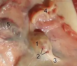
 Ovary of a sheep.
| |
| Details | |
| Drains from | ovary |
| Drains to | inferior vena cava left renal vein |
| Artery | ovarian artery |
| Identifiers | |
| Latin | vena ovarica sinistra, vena ovarica dextra |
| TA98 | A12.3.09.012F A12.3.09.014F |
| TA2 | 5014, 5017 |
| FMA | 14346 |
| Anatomical terminology | |
Structure
It is a paired vein, each one supplying an ovary.
- The right ovarian vein travels through the suspensatory ligament of the ovary and generally joins the inferior vena cava.
- The left ovarian vein, unlike the right, often joins the left renal vein instead of the inferior vena cava.[2][3]
Pathology
Thrombosis of ovarian vein is associated with postpartum endometritis, pelvic inflammatory disease, diverticulitis, appendicitis, and gynecologic surgery.
Additional images
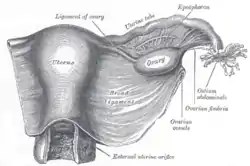
 Uterus and right broad ligament, seen from behind.
See also
References
- II, Anne M.R. Agur, Arthur F. Dalley (2009). Grant's atlas of anatomy. Philadelphia: Wolters Kluwer Health/Lippincott Williams & Wilkins. ISBN 978-0-7817-9604-0.
- Lampmann LE, Smeets AJ, Lohle PN. Uterine fibroids: targeted embolization, an update on technique. Abdom Imaging. 2003 Oct 31; PMID 15160767.
- Drake, Richard L.; Vogl, Wayne; Tibbitts, Adam W.M. Mitchell; illustrations by Richard; Richardson, Paul (2005). Gray's anatomy for students (Pbk. ed.). Philadelphia: Elsevier/Churchill Livingstone. ISBN 978-0-443-06612-2.
External links
- Anatomy photo:40:13-0103 at the SUNY Downstate Medical Center - "Posterior Abdominal Wall: Tributaries to the Inferior Vena Cava"
This article is issued from Wikipedia. The text is licensed under Creative Commons - Attribution - Sharealike. Additional terms may apply for the media files.
