Tunica externa
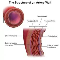
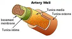
The tunica externa (New Latin "outer coat") — also known as the tunica adventitia (New Latin "additional coat"),[1][2] is the outermost tunica (layer) of a blood vessel, surrounding the tunica media. It is mainly composed of collagen and, in arteries, is supported by external elastic lamina. The collagen serves to anchor the blood vessel to nearby organs, giving it stability.
| Tunica externa | |
|---|---|
 | |
 Section of a medium-sized artery. | |
| Details | |
| Part of | Wall of blood vessels |
| Identifiers | |
| Latin | Tunica externa, tunica adventitia |
| TA98 | A12.0.00.017 |
| TA2 | 3920 |
| TH | H3.09.02.0.01009 |
| Anatomical terminology | |
The three layers of the blood vessels are: an inner tunica intima, a middle tunica media, and an outer tunica externa.
Structure
The tunica externa is made from collagen and elastic fibres in a loose connective tissue.[1][2] This is secreted by fibroblasts.[1]
Function
The tunica externa provides basic structural support to blood vessels.[1] It prevents vessels from expanding too much from internal blood pressure, particularly arteries.[2] It is also relevant in controlling vascular flow in the lungs.[1]
Clinical significance
A common pathological disorder concerning the tunica externa is scurvy, also known as vitamin C deficiency. Scurvy occurs because vitamin C is essential for the synthesis of collagen, and without it, the faulty collagen cannot maintain the vein walls and rupture, leading to a multitude of problems.
Additional images
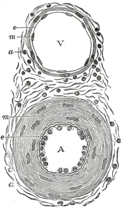 Transverse section through a small artery and vein of the mucous membrane of the epiglottis of a child. (Tunica adventitia is at 'a')
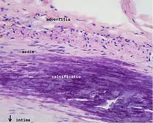 Microphotography of arterial wall with calcified (violet colour) atherosclerotic plaque (haematoxylin & eosin stain)
See also
References
This article incorporates text in the public domain from page 499 of the 20th edition of Gray's Anatomy (1918)
- Hunt, J. M.; Graham, B. B. (2014-01-01), McManus, Linda M.; Mitchell, Richard N. (eds.), "Pulmonary Hypertension/Pulmonary Arterial Hypertension", Pathobiology of Human Disease, San Diego: Academic Press, pp. 2625–2635, doi:10.1016/b978-0-12-386456-7.05306-5, ISBN 978-0-12-386457-4, retrieved 2020-12-29
- Maleszewski, J. J.; Lai, C. K.; Veinot, J. P. (2016-01-01), Buja, L. Maximilian; Butany, Jagdish (eds.), "Chapter 1 - Anatomic Considerations and Examination of Cardiovascular Specimens (Excluding Devices)", Cardiovascular Pathology (Fourth Edition), San Diego: Academic Press, pp. 1–56, doi:10.1016/b978-0-12-420219-1.00001-x, ISBN 978-0-12-420219-1, retrieved 2020-12-29
External links
- Anatomy photo: Circulatory/vessels/vessels7/vessels4 - Comparative Organology at University of California, Davis - "Bird, vessels (LM, High)"
- Image at About.com
| Authority control |
|---|