Protein S
Protein S (also known as S-Protein and spike protein) is a vitamin K-dependent plasma glycoprotein synthesized in the liver. In the circulation, Protein S exists in two forms: a free form and a complex form bound to complement protein C4b-binding protein (C4BP). In humans, protein S is encoded by the PROS1 gene.[5][6]


History
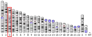
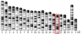
Protein S is named for Seattle, Washington, where it was originally discovered and purified[7] by Earl Davie's group in 1977.[8]
Structure
Protein S is partly homologous to other vitamin K-dependent plasma coagulation proteins, such as protein C and factors VII, IX, and X. Similar to them, it has a Gla domain and several EGF-like domains (four rather than two), but no serine protease domain. Instead, there is a large C-terminus domain that is homologous to plasma steroid hormone-binding proteins such as sex hormone-binding globulin and corticosteroid-binding globulin. It may play a role in the protein functions as either a cofactor for activated protein C (APC) or in binding C4BP.[9][10]
Additionally, protein S has a peptide between the Gla domain and the EGF-like domain, that is cleaved by thrombin. The Gla and EGF-like domains stay connected after the cleavage by a disulfide bond. However, protein S loses its function as an APC cofactor following either this cleavage or binding C4BP.[11]
Function
The best characterized function of Protein S is its role in the anti coagulation pathway, where it functions as a cofactor to Protein C in the inactivation of Factors Va and VIIIa. Only the free form has cofactor activity.[12]
Protein S binds to negatively charged phospholipids via the carboxylated Gla domain. This property allows Protein S to facilitate the removal of cells that are undergoing apoptosis, a form of structured cell death used by the body to remove unwanted or damaged cells. In healthy cells, an ATP (adenosine triphosphate)-dependent enzyme removes negatively charged phospholipids such as phosphatidyl serine from the outer leaflet of the cell membrane. An apoptotic cell (that is, one undergoing apoptosis) no longer actively manages the distribution of phospholipids in its outer membrane and hence begins to display negatively charged phospholipids on its exterior surface. These negatively charged phospholipids are recognized by phagocytes such as macrophages. Protein S binds to the negatively charged phospholipids and functions as a bridge between the apoptotic cell and the phagocyte. This bridging expedites phagocytosis and allows the cell to be removed without giving rise to inflammation or other signs of tissue damage.
Protein S also binds to the nascent complement complex C5,6,7 and prevents this complex from inserting into a membrane. This function prevents the inappropriate activation of the complement system, which would cause uncontrolled systemic inflammation. In fact, Protein S was first discovered in 1977 in this role and it is named after the membrane site that it occupies in the complex.[13]
Pathology
Mutations in the PROS1 gene can lead to Protein S deficiency which is a rare blood disorder which can lead to an increased risk of thrombosis.[14][15]
See also
References
- GRCh38: Ensembl release 89: ENSG00000184500 - Ensembl, May 2017
- GRCm38: Ensembl release 89: ENSMUSG00000022912 - Ensembl, May 2017
- "Human PubMed Reference:". National Center for Biotechnology Information, U.S. National Library of Medicine.
- "Mouse PubMed Reference:". National Center for Biotechnology Information, U.S. National Library of Medicine.
- Lundwall A, Dackowski W, Cohen E, Shaffer M, Mahr A, Dahlbäck B, Stenflo J, Wydro R (September 1986). "Isolation and sequence of the cDNA for human protein S, a regulator of blood coagulation". Proc. Natl. Acad. Sci. U.S.A. 83 (18): 6716–20. Bibcode:1986PNAS...83.6716L. doi:10.1073/pnas.83.18.6716. PMC 386580. PMID 2944113.
- Long GL, Marshall A, Gardner JC, Naylor SL (January 1988). "Genes for human vitamin K-dependent plasma proteins C and S are located on chromosomes 2 and 3, respectively". Somat. Cell Mol. Genet. 14 (1): 93–8. doi:10.1007/BF01535052. PMID 2829367. S2CID 31236887.
- "Protein S deficiency". UpToDate. Retrieved May 10, 2017.
- Kaushansky, K; Lichtman, M; Prchal, J; Levi, M; Press, O; Burns, L; Caligiuri, M (2015). Williams Hematology. McGraw-Hill. p. 1926.
- Stenflo J (1999). "Contributions of Gla and EGF-like domains to the function of vitamin K-dependent coagulation factors". Critical Reviews in Eukaryotic Gene Expression. 9 (1): 59–88. doi:10.1615/CritRevEukaryotGeneExpr.v9.i1.50. PMID 10200912.
- Rosner W (Dec 1991). "Plasma steroid-binding proteins". Endocrinology and Metabolism Clinics of North America. 20 (4): 697–720. doi:10.1016/S0889-8529(18)30240-8. PMID 1778174.
- Dahlbäck B, Lundwall A, Stenflo J (Jun 1986). "Primary structure of bovine vitamin K-dependent protein S". Proceedings of the National Academy of Sciences. 83 (12): 4199–203. Bibcode:1986PNAS...83.4199D. doi:10.1073/pnas.83.12.4199. PMC 323699. PMID 2940598.
- Castoldi E, Hackeng TM (September 2008). "Regulation of coagulation by protein S". Curr. Opin. Hematol. 15 (5): 529–36. doi:10.1097/MOH.0b013e328309ec97. PMID 18695379. S2CID 11522770.
- Podack, Eckhard; Kolb, William; Müller-Eberhard, Hans (1977). "The SC5b-7 complex: formation, isolation, properties, and subunit composition". J. Immunol. 119 (6): 2024–2029. PMID 410885.
- Beauchamp NJ, Dykes AC, Parikh N, Campbell Tait R, Daly ME (June 2004). "The prevalence of, and molecular defects underlying, inherited protein S deficiency in the general population". Br. J. Haematol. 125 (5): 647–54. doi:10.1111/j.1365-2141.2004.04961.x. PMID 15147381. S2CID 705661.
- García de Frutos P, Fuentes-Prior P, Hurtado B, Sala N (September 2007). "Molecular basis of protein S deficiency". Thromb. Haemost. 98 (3): 543–56. doi:10.1160/th07-03-0199. PMID 17849042.
- Heeb, M J; Kojima Y; Rosing J; Tans G; Griffin J H (Dec 1999). "C-terminal residues 621-635 of protein S are essential for binding to factor Va". J. Biol. Chem. UNITED STATES. 274 (51): 36187–92. doi:10.1074/jbc.274.51.36187. ISSN 0021-9258. PMID 10593904. S2CID 45995946.
- Heeb, M J; Mesters R M; Tans G; Rosing J; Griffin J H (Feb 1993). "Binding of protein S to factor Va associated with inhibition of prothrombinase that is independent of activated protein C". J. Biol. Chem. UNITED STATES. 268 (4): 2872–7. doi:10.1016/S0021-9258(18)53854-0. ISSN 0021-9258. PMID 8428962.
Further reading
- Dahlbäck B (1991). "Protein S and C4b-binding protein: components involved in the regulation of the protein C anticoagulant system". Thromb. Haemost. 66 (1): 49–61. doi:10.1055/s-0038-1646373. PMID 1833851.
- Witt, I (2002). "Molekularbiologische Grundlagen und Diagnostik der hereditären Defekte von Antithrombin III, Protein C und Protein S" [Molecular biological basis and diagnosis of hereditary defect of antithrombin III, protein c and protein S]. Hamostaseologie (in German). 22 (2): 14–24. doi:10.1267/Hamo02020057 (inactive 2021-01-14). PMID 12193972. Archived from the original on 2012-03-27. Retrieved 2011-08-19.CS1 maint: DOI inactive as of January 2021 (link)
- Rezende SM, Simmonds RE, Lane DA (2004). "Coagulation, inflammation, and apoptosis: different roles for protein S and the protein S-C4b binding protein complex". Blood. 103 (4): 1192–201. doi:10.1182/blood-2003-05-1551. PMID 12907438. S2CID 133028.
- Dahlbäck B (2007). "The tale of protein S and C4b-binding protein, a story of affection". Thromb. Haemost. 98 (1): 90–6. doi:10.1160/th07-04-0269. PMID 17597997.
- García de Frutos P, Fuentes-Prior P, Hurtado B, Sala N (2007). "Molecular basis of protein S deficiency". Thromb. Haemost. 98 (3): 543–56. doi:10.1160/th07-03-0199. PMID 17849042.
- Maillard C, Berruyer M, Serre CM, et al. (1992). "Protein-S, a vitamin K-dependent protein, is a bone matrix component synthesized and secreted by osteoblasts". Endocrinology. 130 (3): 1599–604. doi:10.1210/en.130.3.1599. PMID 1531628.
- Griffin JH, Gruber A, Fernández JA (1992). "Reevaluation of total, free, and bound protein S and C4b-binding protein levels in plasma anticoagulated with citrate or hirudin". Blood. 79 (12): 3203–11. doi:10.1182/blood.V79.12.3203.bloodjournal79123203. PMID 1534488.
- Guglielmone HA, Vides MA (1992). "A novel functional assay of protein C in human plasma and its comparison with amidolytic and anticoagulant assays". Thromb. Haemost. 67 (1): 46–9. doi:10.1055/s-0038-1648377. PMID 1615482.
- Bertina RM, Ploos van Amstel HK, van Wijngaarden A, et al. (1990). "Heerlen polymorphism of protein S, an immunologic polymorphism due to dimorphism of residue 460". Blood. 76 (3): 538–48. doi:10.1182/blood.V76.3.538.538. PMID 2143091.
- Schmidel DK, Tatro AV, Phelps LG, et al. (1991). "Organization of the human protein S genes". Biochemistry. 29 (34): 7845–52. doi:10.1021/bi00486a010. PMID 2148110.
- Ploos van Amstel HK, Reitsma PH, van der Logt CP, Bertina RM (1991). "Intron-exon organization of the active human protein S gene PS alpha and its pseudogene PS beta: duplication and silencing during primate evolution". Biochemistry. 29 (34): 7853–61. doi:10.1021/bi00486a011. PMID 2148111.
- Allaart CF, Aronson DC, Ruys T, et al. (1991). "Hereditary protein S deficiency in young adults with arterial occlusive disease". Thromb. Haemost. 64 (2): 206–10. PMID 2148653.
- Ohlin AK, Landes G, Bourdon P, et al. (1989). "Beta-hydroxyaspartic acid in the first epidermal growth factor-like domain of protein C. Its role in Ca2+ binding and biological activity". J. Biol. Chem. 263 (35): 19240–8. doi:10.1016/S0021-9258(18)37415-5. PMID 2461936.
- Schwarz HP, Heeb MJ, Lottenberg R, et al. (1989). "Familial protein S deficiency with a variant protein S molecule in plasma and platelets". Blood. 74 (1): 213–21. doi:10.1182/blood.V74.1.213.213. PMID 2526663.
- Ploos van Amstel HK, van der Zanden AL, Reitsma PH, Bertina RM (1987). "Human protein S cDNA encodes Phe-16 and Tyr 222 in consensus sequences for the post-translational processing". FEBS Lett. 222 (1): 186–90. doi:10.1016/0014-5793(87)80217-X. PMID 2820795. S2CID 46365357.
- Dahlbäck B, Lundwall A, Stenflo J (1986). "Primary structure of bovine vitamin K-dependent protein S". Proc. Natl. Acad. Sci. U.S.A. 83 (12): 4199–203. Bibcode:1986PNAS...83.4199D. doi:10.1073/pnas.83.12.4199. PMC 323699. PMID 2940598.
- Lundwall A, Dackowski W, Cohen E, et al. (1986). "Isolation and sequence of the cDNA for human protein S, a regulator of blood coagulation". Proc. Natl. Acad. Sci. U.S.A. 83 (18): 6716–20. Bibcode:1986PNAS...83.6716L. doi:10.1073/pnas.83.18.6716. PMC 386580. PMID 2944113.
- Engesser L, Broekmans AW, Briët E, et al. (1987). "Hereditary protein S deficiency: clinical manifestations". Ann. Intern. Med. 106 (5): 677–82. doi:10.7326/0003-4819-106-5-677. PMID 2952034.
- Watkins PC, Eddy R, Fukushima Y, et al. (1988). "The gene for protein S maps near the centromere of human chromosome 3". Blood. 71 (1): 238–41. doi:10.1182/blood.V71.1.238.238. PMID 2961379.
PDB gallery | |
|---|---|
|
