Round ligament of liver
The round ligament of the liver (or ligamentum teres, or ligamentum teres hepatis) is a ligament that forms part of the free edge of the falciform ligament of the liver. It connects the liver to the umbilicus. It is the remnant of the left umbilical vein. The round ligament divides the left part of the liver into medial and lateral sections.
| Round ligament of liver | |
|---|---|
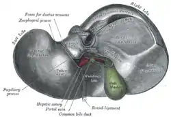 Liver seen from below, with the round ligament labeled at bottom. | |
 1: Right lobe of liver 2: Left lobe of liver 3: Quadrate lobe of liver 4: Round ligament of liver 5: Falciform ligament 6: Caudate lobe of liver 7: Inferior vena cava 8: Common bile duct 9: Hepatic artery 10: Portal vein 11: Cystic duct 12: Hepatic duct 13: Gallbladder | |
| Details | |
| Precursor | left umbilical vein |
| Identifiers | |
| Latin | ligamentum teres hepatis |
| MeSH | D000069592 |
| TA98 | A05.8.01.015 |
| TA2 | 5104 |
| FMA | 14079 |
| Anatomical terminology | |
Structure
The round ligament connects the liver to the umbilicus.[1] It divides the left part of the liver into medial and lateral sections.[2][3]
Development
The round ligament of the liver is the remnant of the left umbilical vein during embryonic development.[1] It only exists in placental mammals.[4] After the child is born, the umbilical vein degenerates to fibrous tissue.[4]
Clinical significance
Portal hypertension
In adulthood, small paraumbilical veins remain in the substance of the ligament. These act as an important portacaval anastomosis in severe portal hypertension, resulting in a caput medusae.[5][6]
Abscess
Very rarely, the round ligament of the liver may develop an abscess. This usually requires liver surgery to treat.[1]
Additional Images
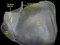 Round ligament of liver.Superior surface of liver.
References
- Sumida, Wataru; Kawashima, Hiroshi; Ishimaru, Tetsuya; Ihara, Yoshiyuki; Kakihara, Tomo; Kato, Reiko; Hayashi, Kentaro; Aoyama, Tomohiro; Omata, Kanako (2019-05-01). "Abscess of ligamentum teres hepatis". Journal of Pediatric Surgery Case Reports. 44: 101198. doi:10.1016/j.epsc.2019.101198. ISSN 2213-5766.
- Gray's anatomy : the anatomical basis of clinical practice. Standring, Susan, (41 ed.). [Philadelphia]. ISBN 978-0-7020-5230-9. OCLC 920806541.CS1 maint: extra punctuation (link) CS1 maint: others (link)
- Moore, Keith L.,. Clinically oriented anatomy. Dalley, Arthur F., II,, Agur, A. M. R., (7 ed.). Philadelphia. ISBN 1-4511-1945-3. OCLC 813301028.CS1 maint: extra punctuation (link) CS1 maint: multiple names: authors list (link)
- Garbar, Veronica; Newton, Bruce W. (2020), "Anatomy, Abdomen and Pelvis, Falciform Ligament", StatPearls, Treasure Island (FL): StatPearls Publishing, PMID 30969680, retrieved 2021-01-24
- Foster, R J; Cowell, G W (2015-04-20). "Acute paraumbilical vein recanalization: an unusual complication of acute pancreatitis". BJR Case Reports. 1 (1): 20150021. doi:10.1259/bjrcr.20150021. ISSN 2055-7159. PMC 6159162. PMID 30363191.
- Mostbeck, G. H.; Wittich, G. R.; Herold, C.; Vergesslich, K. A.; Walter, R. M.; Frotz, S.; Sommer, G. (February 1989). "Hemodynamic significance of the paraumbilical vein in portal hypertension: assessment with duplex US". Radiology. 170 (2): 339–342. doi:10.1148/radiology.170.2.2643137. ISSN 0033-8419. PMID 2643137.
External links
- Anatomy photo:38:12-0106 at the SUNY Downstate Medical Center - "Stomach, Spleen and Liver: The Visceral Surface of the Liver"
- Anatomy image:7819 at the SUNY Downstate Medical Center
- Overview at ucc.edu
- Illustration of Liver Anatomy including ligaments and structures
| Authority control |
|---|