Collagen, type II, alpha 1
Collagen, type II, alpha 1 (primary osteoarthritis, spondyloepiphyseal dysplasia, congenital), also known as COL2A1, is a human gene that provides instructions for the production of the pro-alpha1(II) chain of type II collagen.
Function
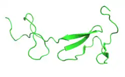
This gene encodes the alpha-1 chain of type II collagen, a fibrillar collagen found in cartilage and the vitreous humor of the eye. Mutations in this gene are associated with achondrogenesis, chondrodysplasia, early onset familial osteoarthritis, SED congenita, Langer-Saldino achondrogenesis, Kniest dysplasia, Stickler syndrome type I, and spondyloepimetaphyseal dysplasia Strudwick type. In addition, defects in processing chondrocalcin, a calcium binding protein that is the C-propeptide of this collagen molecule, are also associated with chondrodysplasia. There are two transcripts identified for this gene.[5] Type II collagen, which adds structure and strength to connective tissues, is found primarily in cartilage, the jelly-like substance that fills the eyeball (the vitreous), the inner ear, and the center portion of the discs between the vertebrae in the spine (nucleus pulposus). Three pro-alpha1(II) chains twist together to form a triple-stranded, ropelike procollagen molecule. These procollagen molecules must be processed by enzymes in the cell. Once these molecules are processed, they leave the cell and arrange themselves into long, thin fibrils that cross-link to one another in the spaces around cells. The cross-linkages result in the formation of very strong mature type II collagen fibers.
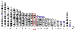
The COL2A1 gene is located on the long (q) arm of chromosome 12 between positions 13.11 and 13.2, from base pair 46,653,017 to base pair 46,684,527.
Related conditions
- Achondrogenesis type 2: Several kinds of mutations in the COL2A1 gene are responsible for achondrogenesis, type 2. These mutations may include missing pieces of the COL2A1 gene, substitution of the amino acid building-block glycine with another amino acid, or changes that cause important parts of the protein to be left out. All of these mutations prevent the normal production of mature type II collagen, which results in achondrogenesis, type 2 by affecting tissues that are rich in type II collagen.
- Platyspondylic lethal skeletal dysplasia, Torrance type:Fewer than 10 mutations in the COL2A1 gene have been identified in people with platyspondylic lethal skeletal dysplasia, Torrance type. Most of these mutations change a single protein building block (amino acid) in the pro-alpha1(II) chain. These COL2A1 mutations lead to the production of an abnormal version of the pro-alpha1(II) chain that cannot be incorporated into type II collagen fibers. As a result, cells make a reduced amount of type II collagen. Instead of forming collagen molecules, the abnormal pro-alpha1(II) chains build up in cartilage cells (chondrocytes). These changes disrupt normal bone development, resulting in skeletal abnormalities such as short arms and legs, a small chest, flattened vertebrae, and short fingers and toes.
- Hypochondrogenesis: Several different types of mutations in the COL2A1 gene are responsible for hypochondrogenesis. These mutations may include missing pieces of the COL2A1 gene, the substitution of the building-block amino acid glycine with another amino acid, or changes that leave out important parts of the protein. All of these changes interfere with the formation of mature triple-stranded type II collagen molecules, which results in this type of hypochondrogenesis by affecting tissues that are rich in type II collagen.
- Kniest dysplasia: Most of the mutations responsible for Kniest dysplasia cause abnormally short pro-alpha1(II) collagen chains to be produced in the cell. These short chains join with longer, normal-length collagen chains. The resulting abnormal type II collagen molecules are shorter than normal, causing the signs and symptoms of Kniest dysplasia.
- Spondyloepimetaphyseal dysplasia, Strudwick type: All of the mutations in the COL2A1 gene characterized to date cause an amino acid switch in the pro-alpha1(II) chain of type II collagen; specifically, the amino acid glycine is replaced by a different amino acid. The substitution of another amino acid for glycine in this chain inhibits the formation of stable, triple-stranded, ropelike collagen molecules. This results in spondyloepimetaphyseal dysplasia, Strudwick type by affecting tissues that are rich in type II collagen.
- Spondyloepiphyseal dysplasia congenita: Spondyloepimetaphyseal dysplasia congenita can be caused by several types of mutations in the COL2A1 gene. These mutations may result in the incorrect substitution of an amino acid in the pro-alpha1(II) chain or the production of an abnormally short pro-alpha1(II) chain. All of these changes interfere with the formation of mature triple-stranded type II collagen molecules, which results in this type of spondyloepimetaphyseal dysplasia congenita by affecting tissues that are rich in type II collagen.
- Spondyloperipheral dysplasia: Mutations that cause spondyloperipheral dysplasia lead to the production of an abnormally short pro-alpha1(II) chain that cannot be incorporated into type II collagen fibers. As a result, cells make a reduced amount of type II collagen. Instead of forming collagen molecules, the abnormal pro-alpha1(II) chains build up in cartilage cells (chondrocytes). These changes disrupt normal bone development, resulting in flattened vertebrae, short fingers and toes, and the other features of spondyloperipheral dysplasia.
- Stickler syndrome: Several of the mutations in the COL2A1 gene result in the production of an abnormally short protein that cannot be incorporated into a type II collagen fiber. Most of the mutations in COL2A1 that cause Stickler syndrome, however, have a premature stop signal in one copy of the gene. Because of this, cells produce only half of the normal amount of pro-alpha 1(II) collagen chains. This shortage results in underproduction of type II collagen in cartilage, causing the symptoms of Stickler syndrome, COL2A1.
- Other disorders with an increased risk from variations of the COL2A1 gene: Variations in the COL2A1 gene may increase the risk of developing osteoarthritis (OA), a degenerative disease of joint cartilage, in some people. The variations in this gene result in amino acid changes in the pro-alpha1(II) chain of type II collagen. These changes in the collagen fibers of the joints are thought to play a role in the wearing down of joint cartilage, resulting in the signs and symptoms of osteoarthritis.
References
- GRCh38: Ensembl release 89: ENSG00000139219 - Ensembl, May 2017
- GRCm38: Ensembl release 89: ENSMUSG00000022483 - Ensembl, May 2017
- "Human PubMed Reference:". National Center for Biotechnology Information, U.S. National Library of Medicine.
- "Mouse PubMed Reference:". National Center for Biotechnology Information, U.S. National Library of Medicine.
- "Entrez Gene: COL2A1 collagen, type II, alpha 1 (primary osteoarthritis, spondyloepiphyseal dysplasia, congenital)".
Further reading
- Chan D, Cole WG, Chow CW, Mundlos S, Bateman JF (January 1995). "A COL2A1 mutation in achondrogenesis type II results in the replacement of type II collagen by type I and III collagens in cartilage". The Journal of Biological Chemistry. 270 (4): 1747–53. doi:10.1074/jbc.270.4.1747. PMID 7829510.
- Cheah KS, Stoker NG, Griffin JR, Grosveld FG, Solomon E (May 1985). "Identification and characterization of the human type II collagen gene (COL2A1)". Proceedings of the National Academy of Sciences of the United States of America. 82 (9): 2555–9. Bibcode:1985PNAS...82.2555C. doi:10.1073/pnas.82.9.2555. PMC 397602. PMID 3857598.
- Donoso LA, Edwards AO, Frost AT, Ritter R, Ahmad N, Vrabec T, Rogers J, Meyer D, Parma S (2003). "Clinical variability of Stickler syndrome: role of exon 2 of the collagen COL2A1 gene". Survey of Ophthalmology. 48 (2): 191–203. doi:10.1016/S0039-6257(02)00460-5. PMID 12686304.
- Fernandes RJ, Wilkin DJ, Weis MA, Wilcox WR, Cohn DH, Rimoin DL, Eyre DR (July 1998). "Incorporation of structurally defective type II collagen into cartilage matrix in kniest chondrodysplasia". Archives of Biochemistry and Biophysics. 355 (2): 282–90. doi:10.1006/abbi.1998.0745. PMID 9675039.
- Ikeda T, Mabuchi A, Fukuda A, Kawakami A, Ryo Y, Yamamoto S, Miyoshi K, Haga N, Hiraoka H, Takatori Y, Kawaguchi H, Nakamura K, Ikegawa S (July 2002). "Association analysis of single nucleotide polymorphisms in cartilage-specific collagen genes with knee and hip osteoarthritis in the Japanese population". Journal of Bone and Mineral Research. 17 (7): 1290–6. doi:10.1359/jbmr.2002.17.7.1290. PMID 12096843. S2CID 31291501.
- Körkkö J, Cohn DH, Ala-Kokko L, Krakow D, Prockop DJ (May 2000). "Widely distributed mutations in the COL2A1 gene produce achondrogenesis type II/hypochondrogenesis". American Journal of Medical Genetics. 92 (2): 95–100. doi:10.1002/(SICI)1096-8628(20000515)92:2<95::AID-AJMG3>3.0.CO;2-9. PMID 10797431.
- Meulenbelt I, Bijkerk C, De Wildt SC, Miedema HS, Breedveld FC, Pols HA, Hofman A, Van Duijn CM, Slagboom PE (September 1999). "Haplotype analysis of three polymorphisms of the COL2A1 gene and associations with generalised radiological osteoarthritis". Annals of Human Genetics. 63 (Pt 5): 393–400. doi:10.1046/j.1469-1809.1999.6350393.x. PMID 10735581. S2CID 21918163.
- Mortier GR, Weis M, Nuytinck L, King LM, Wilkin DJ, De Paepe A, Lachman RS, Rimoin DL, Eyre DR, Cohn DH (April 2000). "Report of five novel and one recurrent COL2A1 mutations with analysis of genotype-phenotype correlation in patients with a lethal type II collagen disorder". Journal of Medical Genetics. 37 (4): 263–71. doi:10.1136/jmg.37.4.263. PMC 1734564. PMID 10745044.
- Richards AJ, Baguley DM, Yates JR, Lane C, Nicol M, Harper PS, Scott JD, Snead MP (November 2000). "Variation in the vitreous phenotype of Stickler syndrome can be caused by different amino acid substitutions in the X position of the type II collagen Gly-X-Y triple helix". American Journal of Human Genetics. 67 (5): 1083–94. doi:10.1016/S0002-9297(07)62938-3. PMC 1288550. PMID 11007540.
- Snead MP, Yates JR (May 1999). "Clinical and Molecular genetics of Stickler syndrome". Journal of Medical Genetics. 36 (5): 353–9. doi:10.1136/jmg.36.5.353 (inactive 2021-01-14). PMC 1734362. PMID 10353778.CS1 maint: DOI inactive as of January 2021 (link)
- Tiller GE, Polumbo PA, Weis MA, Bogaert R, Lachman RS, Cohn DH, Rimoin DL, Eyre DR (September 1995). "Dominant mutations in the type II collagen gene, COL2A1, produce spondyloepimetaphyseal dysplasia, Strudwick type". Nature Genetics. 11 (1): 87–9. doi:10.1038/ng0995-87. PMID 7550321. S2CID 24826973.
- Tysoe C, Saunders J, White L, Hills N, Nicol M, Evans G, Cole T, Chapman S, Pope FM (September 2003). "A glycine to aspartic acid substitution of COL2A1 in a family with the Strudwick variant of spondyloepimetaphyseal dysplasia". QJM. 96 (9): 663–71. doi:10.1093/qjmed/hcg112. PMID 12925722.
- Weis MA, Wilkin DJ, Kim HJ, Wilcox WR, Lachman RS, Rimoin DL, Cohn DH, Eyre DR (February 1998). "Structurally abnormal type II collagen in a severe form of Kniest dysplasia caused by an exon 24 skipping mutation". The Journal of Biological Chemistry. 273 (8): 4761–8. doi:10.1074/jbc.273.8.4761. PMID 9468540.
- Wilkin DJ, Artz AS, South S, Lachman RS, Rimoin DL, Wilcox WR, McKusick VA, Stratakis CA, Francomano CA, Cohn DH (July 1999). "Small deletions in the type II collagen triple helix produce kniest dysplasia". American Journal of Medical Genetics. 85 (2): 105–12. doi:10.1002/(SICI)1096-8628(19990716)85:2<105::AID-AJMG2>3.0.CO;2-Z. PMID 10406661.
- Zabel B, Hilbert K, Stöss H, Superti-Furga A, Spranger J, Winterpacht A (May 1996). "A specific collagen type II gene (COL2A1) mutation presenting as spondyloperipheral dysplasia". American Journal of Medical Genetics. 63 (1): 123–8. doi:10.1002/(SICI)1096-8628(19960503)63:1<123::AID-AJMG22>3.0.CO;2-P. PMID 8723097.
- Zankl A, Neumann L, Ignatius J, Nikkels P, Schrander-Stumpel C, Mortier G, Omran H, Wright M, Hilbert K, Bonafé L, Spranger J, Zabel B, Superti-Furga A (February 2005). "Dominant negative mutations in the C-propeptide of COL2A1 cause platyspondylic lethal skeletal dysplasia, torrance type, and define a novel subfamily within the type 2 collagenopathies". American Journal of Medical Genetics Part A. 133A (1): 61–7. doi:10.1002/ajmg.a.30531. PMID 15643621. S2CID 24925027.
- Zankl A, Zabel B, Hilbert K, Wildhardt G, Cuenot S, Xavier B, Ha-Vinh R, Bonafé L, Spranger J, Superti-Furga A (August 2004). "Spondyloperipheral dysplasia is caused by truncating mutations in the C-propeptide of COL2A1". American Journal of Medical Genetics Part A. 129A (2): 144–8. doi:10.1002/ajmg.a.30222. PMID 15316962. S2CID 22472125.


