Monoblast
Monoblasts are the committed progenitor cells that differentiated from myeloid stem cell in the process of hematopoiesis. Their myeloid cell fate is induced by the concentration of transcription factors they are surrounded by during development.[1] They are normally found in bone marrow and do not appear in the normal peripheral blood.[2] They mature into monocytes which, in turn, develop into macrophages.[3]
| Monoblast | |
|---|---|
 Monoblast | |
| Details | |
| System | Immune system |
| Location | Bone marrow |
| Function | A precursor monocyte |
| Identifiers | |
| TH | H2.00.04.3.08002 |
| Anatomical terms of microanatomy | |
Structure
A typical monoblast is about 12 to 20 μm in diameter, has a nuclear to cytoplasm ratio of 4:1 to 3:1, and, like most myeloid blasts, has a round to oval nucleus with fine chromatin structure. One to four nucleoli are usually visible. The nucleus can be central or eccentric and it can show evidence of indentation or folding. The cytoplasm stains moderately to lightly basophilic and may contain small azurophilic granules.[3][4]
Development
The monoblast is the first stage of monocyte-macrophage maturation. The developmental stages of the monoblast are: CFU-GM (pluripotential hemopoietic stem cell or hemocytoblast) -> monoblast -> promonocyte -> monocyte-> macrophage.[3][5]
Additional images
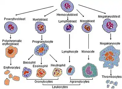 Blood cell lineage
_diagram_en.svg.png.webp) Hematopoiesis
Hematopoiesis
References
- Punt, Jenni; Stranford, Sharon A.; Jones, Patricia P.; Owen, Judith A. Kuby Immunology (Eighth ed.). New York: MacMillian. ISBN 978-1-4641-8978-4. OCLC 1002672752.
- Abbas, Abul K.; Lichtman, Andrew H.; Pillai, Shiv (2012). Cellular and molecular immunology (7th ed.). Philadelphia: Elsevier/Saunders. ISBN 9781437715286. OCLC 698580696.
- Mescher, Anthony L.; Junqueira, Luiz Carlos Uchôa (2013-02-22). Junqueira's basic histology: text and atlas (Thirteenth ed.). New York: McGraw-Hill Medical. ISBN 9780071807203. OCLC 854567882.
- Glassy, Eric F. (2018). Color atlas of hematology: an illustrated field guide based on proficiency testing: peripheral blood (Second ed.). Northfield, Illinois: College of American Pathologists. ISBN 9781941096390. OCLC 1023810216.
- Ross, Michael H.; Pawlina, Wojciech (2011). Histology – a text and atlas – with correlated cell and molecular biology (6th ed.). Philadelphia: Wolters Kluwer/Lippincott Williams & Wilkins Health. ISBN 9780781772006. OCLC 548651322.