Green sulfur bacteria
The green sulfur bacteria (Chlorobiaceae) are a family of obligately anaerobic photoautotrophic bacteria. Together with the non-photosynthetic Ignavibacteriaceae, they form the phylum Chlorobi.[1]
| Green sulfur bacteria | |
|---|---|
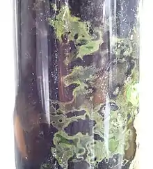 | |
| Green sulfur bacteria in a Winogradsky column | |
| Scientific classification | |
| Domain: | |
| Superphylum: | |
| (unranked): | |
| Phylum: | Chlorobi Iino et al. 2010 |
| Genus | |
| |
Green sulfur bacteria are nonmotile (except Chloroherpeton thalassium, which may glide) and capable of anoxygenic photosynthesis.[1][2] In contrast to plants, green sulfur bacteria mainly use sulfide ions as electron donors.[3] They are autotrophs that utilize the reverse tricarboxylic acid cycle to perform carbon fixation.[4] Green sulfur bacteria have been found in depths of up to 145m in the Black Sea, with low light availability.[5]
Characteristics of green-Sulfur bacteria:
Major photosynthetic pigment: Bacteriochlorophylls a plus c, d or e
Location of photosynthetic pigment: Chlorosomes and plasma membranes
Photosynthetic electron donor: H2, H2S, S
Sulfur deposition: Outside of the cell
Metabolic type: Photolithoautotrophs[6]
Habitat
The Black Sea, an extremely anoxic environment, was found to house a large population of green sulfur bacteria at about 100 m depth. Due to the lack of light available in this region of the sea, most bacteria were photosynthetically inactive. The photosynthetic activity detected in the sulfide chemocline suggests that the bacteria need very little energy for cellular maintenance.[5]
A species of green sulfur bacteria has been found living near a black smoker off the coast of Mexico at a depth of 2,500 m in the Pacific Ocean. At this depth, the bacterium, designated GSB1, lives off the dim glow of the thermal vent since no sunlight can penetrate to that depth.[7]
Phylogeny
The currently accepted phylogeny is based on 16S rRNA-based LTP release 123 by The All-Species Living Tree Project.[8]
| |||||||||||||||||||||||||||||||||||||||||||||||||||||||||||||||||||||||||
Taxonomy
The currently accepted taxonomy is based on the List of Prokaryotic names with Standing in Nomenclature.(LSPN)[9][10]
- Phylum Chlorobi Iino et al. 2010
- Class Ignavibacteria Iino et al. 2010
- Order Ignavibacteriales Iino et al. 2010
- Family Ignavibacteriaceae Iino et al. 2010
- Genus Ignavibacterium Iino et al. 2010 emend. Podosokorskaya et al. 2013
- Species Ignavibacterium album Iino et al. 2010 emend. Podosokorskaya et al. 2013
- Genus Melioribacter roseus Podosokorskaya et al. 2013 ["Melioribacter" Podosokorskaya et al. 2011]
- Species Melioribacter roseus Podosokorskaya et al. 2011 ["Melioribacter roseus" Podosokorskaya et al. 2011]
- Genus Ignavibacterium Iino et al. 2010 emend. Podosokorskaya et al. 2013
- Family Ignavibacteriaceae Iino et al. 2010
- Order Ignavibacteriales Iino et al. 2010
- Class Chlorobea Cavalier-Smith 2002
- Order Chlorobiales Gibbons and Murray 1978
- Family Chlorobiaceae Copeland 1956
- Genus Ancalochloris Gorlenko and Lebedeva 1971
- Species Ancalochloris perfilievii[notes 3]Gorlenko and Lebedeva 1971
- Genus Chlorobaculum Imhoff 2003
- Species "C. macestae"[notes 1]Keppen et al. 2008
- Species C. limnaeum Imhoff 2003
- Species C. parvum Imhoff 2003
- Species C. tepidum (Wahlund et al. 1996) Imhoff 2003 (type sp.) ["Chlorobium tepidum" Wahlund et al. 1991; Chlorobium tepidum Wahlund et al. 1996]
- Species C. thiosulfatiphilum Imhoff 2003 ["Chlorobium limicola f. sp. thiosulfatophilum" (Larsen 1952) Pfennig & Truper 1971]
- Genus Chlorobium Nadson 1906 emend. Imhoff 2003
- Species Chlorobium chlorovibrioides[notes 2](Gorlenko et al. 1974) Imhoff 2003
- Species C. bathyomarinum[notes 1][7]
- Species C. chlorochromatii[notes 1]Vogl et al. 2006 (epibiont of the phototrophic consortium Chlorochromatium aggregatum) ["Chlorobium chlorochromatii" Meschner 1957]
- Species C. gokarna[notes 1]Anil Kumar 2005
- Species C. clathratiforme (Szafer 1911) emend. Imhoff 2003 ["Aphanothece clathratiformis" Szafer 1911; "Pelodictyon lauterbornii" Geitler 1925; Pelodictyon clathratiforme (Szafer 1911) Lauterborn 1913]
- Species C. ferrooxidans Heising et al. 1998 emend. Imhoff 2003
- Species C. luteolum (Schmidle 1901) emend. Imhoff 2003 ["Aphanothece luteola" Schmidle 1901; "Pelodictyon aggregatum" Perfil'ev 1914; "Schmidlea luteola" (Schmidle 1901) Lauterborn 1913; Pelodictyon luteolum (Schmidle 1901) Pfennig and Truper 1971]
- Species C. limicola Nadson 1906 emend. Imhoff 2003 (type sp.)
- Species C. phaeobacteroides Pfennig 1968 emend. Imhoff 2003
- Species C. phaeovibrioides Pfennig 1968 emend. Imhoff 2003
- Genus Chloroherpeton Gibson et al. 1985
- Species Chloroherpeton thalassium Gibson et al. 1985
- Genus Clathrochloris Witt et al. 1989
- Species "Clathrochloris sulfurica"[notes 1]Witt et al. 1989
- Genus Pelodictyon Lauterborn 1913
- Species Pelodictyon phaeum Gorlenko 1972
- Genus Prosthecochloris Gorlenko 1970 emend. Imhoff 2003
- Genus Ancalochloris Gorlenko and Lebedeva 1971
- Family Chlorobiaceae Copeland 1956
- Order Chlorobiales Gibbons and Murray 1978
Notes
Metabolism
Photosynthesis in the green sulfur bacteria
The green sulfur bacteria use a Type I reaction center for photosynthesis. Type I reaction centers are the bacterial homologue of photosystem I (PSI) in plants and cyanobacteria. The GSB reaction centers contain bacteriochlorophyll a and are known as P840 reaction centers due to the excitation wavelength of 840 nm that powers the flow of electrons. In green sulfur bacteria the reaction center is associated with a large antena complex called the chlorosome that captures and funnels light energy to the reaction center. The chlorosomes have a peak absorption in the far red region of the spectrum between 720-750 nm because they contain bacteriocholorophyll c, d and e.[11] A protein complex called the Fenna-Matthews-Olson complex (FMO) is physically located between the chlorosomes and the P840 RC. The FMO complex helps efficiently transfer the energy absorbed by the antena to the reaction center.
PSI and Type I reaction centers are able to reduce ferredoxin (Fd), a strong reductant that can be used to fix CO
2 and reduce NADPH. Once the reaction center (RC) has given an electron to Fd it becomes an oxidizing agent (P840+) with a reduction potential of around +300 mV. While this is not positive enough to strip electrons from water to synthesize O
2 (E
0 = +820 mV), it can accept electrons from other sources like H
2S, thiosulphate or Fe2+
ions.[12] This transport of electrons from donors like H
2S to the acceptor Fd is called linear electron flow or linear electron transport. The oxidation of sulfide ions leads to the production of sulfur as a waste product that accumulates as globules on the extracellular side of the membrane.These globules of sulfur give green sulfur bacteria their name. When sulfide is depleted, the sulfur globules are consumed and further oxidized to sulfate. However, the pathway of sulfur oxidation is not well-understood.[3]
Instead of passing the electrons onto Fd, the Fe-S clusters in the P840 reaction center can transfer the electrons to menaquinone (MQ:MQH
2) which returns the electrons to the P840+ via an electron transport chain (ETC). On the way back to the RC the electrons from MQH2 pass through a cytochrome bc1 complex (similar to the complex III of mitochondria) that pumps H+
ions across the membrane. The electrochemical potential of the protons across the membrane is used to synthesize ATP by the FoF1 ATP synthase. This cyclic electron transport is responsible for converting light energy into cellular energy in the form of ATP.
Carbon fixation of green sulfur bacteria
Green sulfur bacteria are photoautotrophs: they not only get energy from light, they can grow using carbon dioxide as their sole source of carbon. They fix carbon dioxide using the reverse tricarboxylic acid cycle (rTCA) cycle[4] where energy is consumed to reduce carbon dioxide in order to synthesize pyruvate and acetate. These molecules are used as the raw materials to synthesize all the building blocks a cell needs to generate macromolecules. The rTCA cycle is highly energy efficient enabling the bacteria to grow under low light conditions.[13] However it has several oxygen sensitive enzymes that limits it's efficiency in aerobic conditions.[13]
The reactions of reversal of the oxidative tricarboxylic acid cycle are catalyzed by four enzymes:[4]
- pyruvate:ferredoxin (Fd) oxidoreductase:
- acetyl-CoA + CO2 + 2Fdred + 2H+ ⇌ pyruvate + CoA + 2Fdox
- ATP citrate lyase:
- ACL, acetyl-CoA + oxaloacetate + ADP + Pi ⇌ citrate + CoA + ATP
- α-keto-glutarate:ferredoxin oxidoreductase:
- succinyl-CoA + CO2 + 2Fdred + 2H+ ⇌ α-ketoglutarate + CoA + 2Fdox
- fumarare reductase
- succinate + acceptor ⇌ fumarate + reduced acceptor
Mixotrophy in green sulfur bacteria
Green sulfur bacteria are obligate photoautotrophs: they cannot grow in the absence of light even if they are provided with organic matter.[4][12] However they exhibit a form of mixotrophy where they can consume simple organic compounds in the presence of light and CO2.[4]
Nitrogen fixation
The majority of green sulfur bacteria are diazotrophs: they can reduce nitrogen to ammonia which is then used to synthesize amino acids.[14]
References
- Bryant DA, Frigaard NU (November 2006). "Prokaryotic photosynthesis and phototrophy illuminated". Trends in Microbiology. 14 (11): 488–96. doi:10.1016/j.tim.2006.09.001. PMID 16997562.
- Green BR (2003). Light-Harvesting Antennas in Photosynthesis. p. 8. ISBN 0792363353.
- Sakurai H, Ogawa T, Shiga M, Inoue K (June 2010). "Inorganic sulfur oxidizing system in green sulfur bacteria". Photosynthesis Research. 104 (2–3): 163–76. doi:10.1007/s11120-010-9531-2. PMID 20143161. S2CID 1091791.
- Tang KH, Blankenship RE (November 2010). "Both forward and reverse TCA cycles operate in green sulfur bacteria". The Journal of Biological Chemistry. 285 (46): 35848–54. doi:10.1074/jbc.M110.157834. PMC 2975208. PMID 20650900.
- Marschall E, Jogler M, Hessge U, Overmann J (May 2010). "Large-scale distribution and activity patterns of an extremely low-light-adapted population of green sulfur bacteria in the Black Sea". Environmental Microbiology. 12 (5): 1348–62. doi:10.1111/j.1462-2920.2010.02178.x. PMID 20236170.
- Pranav kumar, Usha mina (2014). Life science fundamental and practice part I.
- Beatty JT, Overmann J, Lince MT, Manske AK, Lang AS, Blankenship RE, Van Dover CL, Martinson TA, Plumley FG (June 2005). "An obligately photosynthetic bacterial anaerobe from a deep-sea hydrothermal vent". Proceedings of the National Academy of Sciences of the United States of America. 102 (26): 9306–10. Bibcode:2005PNAS..102.9306B. doi:10.1073/pnas.0503674102. PMC 1166624. PMID 15967984.
- See the All-Species Living Tree Project . Data extracted from the "16S rRNA-based LTP release 123 (full tree)" (PDF). Silva Comprehensive Ribosomal RNA Database. Retrieved 2016-03-20.
- See the List of Prokaryotic names with Standing in Nomenclature. Data extracted from J.P. Euzéby. "Chlorobi". Archived from the original on 2013-01-27. Retrieved 2016-03-20.
- See the NCBI webpage on Chlorobi Data extracted from Sayers; et al. "NCBI Taxonomy Browser". National Center for Biotechnology Information. Retrieved 2016-03-20.
- Hauska G, Schoedl T, Remigy H, Tsiotis G (October 2001). "The reaction center of green sulfur bacteria(1)". Biochimica et Biophysica Acta. 1507 (1–3): 260–77. doi:10.1016/S0005-2728(01)00200-6. PMID 11687219.
- Ligrone, Roberto (2019). "Moving to the Light: The Evolution of Photosynthesis". In Roberto Ligrone (ed.). Biological Innovations that Built the World: A Four-billion-year Journey through Life and Earth History. Cham: Springer International Publishing. pp. 99–127. doi:10.1007/978-3-030-16057-9_4. ISBN 978-3-030-16057-9. Retrieved 2021-01-29.
- Bar-Even, Arren; Noor, Elad; Milo, Ron (2012). "A survey of carbon fixation pathways through a quantitative lens". Journal of Experimental Botany. 63 (6): 2325–2342. doi:10.1093/jxb/err417. ISSN 1460-2431.
- Madigan, Michael T. (1995). "Microbiology of Nitrogen Fixation by Anoxygenic Photosynthetic Bacteria". In Robert E. Blankenship; Michael T. Madigan; Carl E. Bauer (eds.). Anoxygenic Photosynthetic Bacteria. Advances in Photosynthesis and Respiration. Dordrecht: Springer Netherlands. pp. 915–928. doi:10.1007/0-306-47954-0_42. ISBN 978-0-306-47954-0.
External links
- "The Family Chlorobiaceae". The Prokaryotes. Archived from the original on November 17, 2003. Retrieved July 5, 2005.