Myostatin
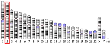
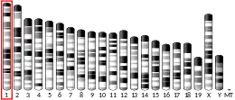
Myostatin (also known as growth differentiation factor 8, abbreviated GDF-8) is a myokine, a protein produced and released by myocytes that acts on muscle cells' autocrine function to inhibit myogenesis: muscle cell growth and differentiation. In humans it is encoded by the MSTN gene.[6] Myostatin is a secreted growth differentiation factor that is a member of the TGF beta protein family.[7][8]

Animals either lacking myostatin or treated with substances that block the activity of myostatin have significantly more muscle mass. Furthermore, individuals who have mutations in both copies of the myostatin gene have significantly more muscle mass and are stronger than normal. There is hope that studies into myostatin may have therapeutic application in treating muscle wasting diseases such as muscular dystrophy.[9]
Discovery and sequencing
The gene encoding myostatin was discovered in 1997 by geneticists Se-Jin Lee and Alexandra McPherron who produced a knockout strain of mice that lack the gene, and have approximately twice as much muscle as normal mice.[10] These mice were subsequently named "mighty mice".
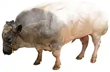
Naturally occurring deficiencies of myostatin of various sorts have been identified in some breeds of cattle,[11] sheep,[12] whippets,[13] and humans.[14] In each case the result is a dramatic increase in muscle mass.
Structure and mechanism of action
Human myostatin consists of two identical subunits, each consisting of 109 (NCBI database claims human myostatin is 375 residues long) amino acid residues [note the full length gene encodes a 375AA prepro-protein which is proteolytically processed to its shorter active form].[15][16] Its total molecular weight is 25.0 kDa. The protein is inactive until a protease cleaves the NH2-terminal, or "pro-domain" portion of the molecule, resulting in the active COOH-terminal dimer. Myostatin binds to the activin type II receptor, resulting in a recruitment of either coreceptor Alk-3 or Alk-4. This coreceptor then initiates a cell signaling cascade in the muscle, which includes the activation of transcription factors in the SMAD family—SMAD2 and SMAD3. These factors then induce myostatin-specific gene regulation. When applied to myoblasts, myostatin inhibits their differentiation into mature muscle fibers.
Myostatin also inhibits Akt, a kinase that is sufficient to cause muscle hypertrophy, in part through the activation of protein synthesis. However, Akt is not responsible for all of the observed muscle hyperthrophic effects which are mediated by myostatin inhibition[17] Thus myostatin acts in two ways: by inhibiting muscle differentiation, and by inhibiting Akt-induced protein synthesis.
Effects in animals
Double muscled cattle

After that discovery, several laboratories cloned and established the nucleotide sequence of a myostatin gene in two breeds of cattle, Belgian Blue and Piedmontese. They found mutations in the myostatin gene (various mutations in each breed) which in one way or another lead to absence of functional myostatin.[10][11][18] Unlike mice with a damaged myostatin gene, in these cattle breeds the muscle cells multiply rather than enlarge. People describe these cattle breeds as "double muscled", but the total increase in all muscles is no more than 40%.[11][19][20]
Animals lacking myostatin or animals treated with substances such as follistatin that block the binding of myostatin to its receptor have significantly larger muscles. Thus, reduction of myostatin could potentially benefit the livestock industry, with even a 20 percent reduction in myostatin levels potentially having a large effect on the development of muscles.[21]
However, the animal breeds developed as homozygous for myostatin deficiency have reproduction issues due to their unusually heavy and bulky offspring, and require special care and a more expensive diet to achieve a superior yield. This negatively affects economics of myostatin-deficient breeds to the point where they do not usually offer an obvious advantage. While hypertrophic meat (e.g. from Piedmontese beef) has a place on the specialist market due to its unusual properties, at least for purebred myostatin-deficient strains the expenses and (especially in cattle) necessity of veterinary supervision place them at a disadvantage in the bulk market.[22]
Whippets

Whippets can have a mutation of the myostatin which involves a two-base-pair deletion, and results in a truncated, and likely inactive, myostatin protein.
Animals with a homozygous deletion have an unusual body shape, with a broader head, pronounced overbite, shorter legs, and thicker tails, and are called "bully whippets" by the breeding community. Although significantly more muscular, they are less able runners than other whippets. However, whippets that were heterozygous for the mutation were significantly over-represented in the top racing classes.[13]
Rabbits and goats
In 2016, the CRISPR/Cas9 system was used to genetically engineer rabbits and goats with no functional copies of the myostatin gene.[23] In both cases the resulting animals were significantly more muscular. However, rabbits without myostatin also exhibited an enlarged tongue, a higher rate of still births, and a reduced lifespan.
Pigs
A South Korean-Chinese team has engineered "double muscle" pigs, as with cattle, aiming for cheaper breeds for the meat market.[24] Similar health problems have resulted as with other mammals, such as birthing difficulties due to excessive size.[24]
Clinical significance
Mutations
A technique for detecting mutations in myostatin variants has been developed.[25] Mutations that reduce the production of functional myostatin lead to an overgrowth of muscle tissue. Myostatin-related muscle hypertrophy has an incomplete autosomal dominance pattern of inheritance. People with a mutation in both copies of the MSTN gene in each cell (homozygotes) have significantly increased muscle mass and strength. People with a mutation in one copy of the MSTN gene in each cell (heterozygotes) have increased muscle bulk, but to a lesser degree.
In humans
In 2004, a German boy was diagnosed with a mutation in both copies of the myostatin-producing gene, making him considerably stronger than his peers. His mother has a mutation in one copy of the gene.[14][26][27]
An American boy born in 2005 was diagnosed with a clinically similar condition, but with a somewhat different cause:[28] his body produces a normal level of functional myostatin, but because he is stronger and more muscular than most others his age, a defect in his myostatin receptors is thought to prevent his muscle cells from responding normally to myostatin. He appeared on the television show World's Strongest Toddler.[29]
Therapeutic potential
Further research into myostatin and the myostatin gene may lead to therapies for muscular dystrophy.[9][30] The idea is to introduce substances that block myostatin. A monoclonal antibody specific to myostatin increases muscle mass in mice[31] and monkeys.[21]
A two-week treatment of normal mice with soluble activin type IIB receptor, a molecule that is normally attached to cells and binds to myostatin, leads to a significantly increased muscle mass (up to 60%).[32] It is thought that binding of myostatin to the soluble activin receptor prevents it from interacting with the cell-bound receptors. In September 2020 scientists reported that suppressing activin type 2 receptors-signalling proteins myostatin and activin A via activin A/myostatin inhibitor ACVR2B – tested preliminarily in humans in the form of ACE-031 in the early 2010s[33][34] – can protect against both muscle and bone loss in mice. The mice were sent to the International Space Station and could largely maintain their muscle weights – about twice those of wild type due to genetic engineering for targeted deletion of the myostatin gene – under microgravity.[35][36] Treating progeric mice with soluble activin receptor type IIB before the onset of premature ageing signs appear to protects against muscle loss and delay age related signs in other organs.[37]
It remains unclear as to whether long-term treatment of muscular dystrophy with myostatin inhibitors is beneficial, as the depletion of muscle stem cells could worsen the disease later on. As of 2012, no myostatin-inhibiting drugs for humans are on the market. An antibody genetically engineered to neutralize myostatin, stamulumab, which was under development by pharmaceutical company Wyeth,[38] is no longer under development.[39] Some athletes, eager to get their hands on such drugs, turn to the internet where fake "myostatin blockers" are being sold.[21]
Myostatin levels are effectively decreased by creatine supplementation.[40]
Myostatin levels can be temporarily reduced using a cholesterol-conjugated siRNA gene knockdown.[41]
Athletic use
Inhibition of myostatin leads to muscle hyperplasia and hypertrophy. Myostatin inhibitors can improve athletic performance and therefore there is a concern these inhibitors might be abused in the field of sports.[42] However, studies in mice suggest that myostatin inhibition does not directly increase the strength of individual muscle fibers.[43] Myostatin inhibitors are specifically banned by the World Anti-Doping Agency (WADA).[44] In an August 12, 2012, interview with National Public Radio, Carlon Colker stated "when the myostatin inhibitors come along, they'll be abused. There's no question in my mind."[45]
Effects
On bone formation
Due to myostatin's ability to inhibit muscle growth, it can indirectly inhibit bone formation by decreasing the load on the bone.[46][47] It has a direct signalling effect on bone formation[48] as well as degradation.[49][47] Knockdown of myostatin has been shown to reduce formation of osteoclasts (multinucleated cells responsible for the breakdown of bone tissue) in mice modeling rheumatoid arthritis.[49] Rheumatoid arthritis is an autoimmune disorder that, among other effects, leads to the degradation of the bone tissue in affected joints. Myostatin has not, however, been shown to be solely sufficient for the formation of mature osteoclasts from macrophages, only an enhancer.
Myostatin expression is increased around the site of a fracture. Suppression of myostatin at the fracture site leads to increased callus and overall bone size, further supporting the inhibitory effect of myostatin on bone formation. One study[49] by Berno Dankbar et al., 2015 found that myostatin deficiency leads to a notable reduction in inflammation around a fracture site. Myostatin affects osteoclastogenesis by binding to receptors on osteoclastic macrophages and causing a signalling cascade. The downstream signalling cascade enhances the expression of RANKL-dependent integrin αvβ3, DC-STAMP, calcitonin receptors, and NFATc1 (which is part of the initial intracellular complex that starts the signaling cascade, along with R-Smad2 and ALK4 or ALK5).[49][47]
An association between osteoporosis, another disease characterized by the degradation of bony tissue, and sarcopenia, the age-related degeneration of muscle mass and quality have also been found.[47] Whether this link is a result of direct regulation or a secondary effect through muscle mass is not known.
A link in mice between the concentration of myostatin in the prenatal environment and the strength of offspring's bones, partially counteracting the effects of osteogenesis imperfecta (brittle bone disease) has been found.[50] Osteogenesis imperfecta is due to a mutation that causes the production of abnormal Type I collagen. Mice with defective myostatin were created by replacing sequences coding for the C-terminal region of myostatin with a neomycin cassette, rendering the protein nonfunctional. By crossbreeding mice with the abnormal Type I collagen and those with the knockout myostatin, the offspring had "a 15% increase in torsional ultimate strength, a 29% increase in tensile strength, and a 24% increase in energy to failure" of their femurs as compared to the other mice with osteogenesis imperfecta, showing the positive effects of decreased myostatin on bone strength and formation.[51]
On the heart
Myostatin is expressed at very low levels in cardiac myocytes.[52][53] Although its presence has been noted in cardiomyocytes of both fetal and adult mice,[54] its physiological function remains uncertain.[53] However, it has been suggested that fetal cardiac myostatin may play a role in early heart development.[54]
Myostatin is produced as promyostatin, a precursor protein kept inactive by the latent TGF-β binding protein 3 (LTBP3).[52] Pathological cardiac stress promotes N-terminal cleavage by furin convertase to create a biologically active C-terminal fragment. The mature myostatin is then segregated from the latent complex via proteolytic cleavage by BMP-1 and tolloid metallopreoteinases.[52] Free myostatin is able to bind its receptor, ActRIIB, and increase SMAD2/3 phosphorylation.[52] The latter produces a heteromeric complex with SMAD4, inducing myostatin translocation into the cardiomyocyte nucleus to modulate transcription factor activity.[55] Manipulating the muscle creatinine kinase promoter can modulate myostatin expression, although it has only been observed in male mice thus far.[52][53]
Myostatin may inhibit cardiomyocyte proliferation and differentiation by manipulating cell cycle progression.[54] This argument is supported by the fact that myostatin mRNA is poorly expressed in proliferating fetal cardiomyocytes.[52][55] In vitro studies indicate that myostatin promotes SMAD2 phosphorylation to inhibit cardiomyocyte proliferation. Furthermore, myostatin has been shown to directly prevent cell cycle G1 to S phase transition by decreasing levels of cyclin-dependent kinase complex 2 (CDK2) and by increasing p21 levels.[55]
Growth of cardiomyocytes may also be hindered by myostatin-regulated inhibition of protein kinase p38 and the serine-threonine protein kinase Akt, which typically promote cardiomyocyte hypertrophy.[56] However, increased myostatin activity only occurs in response to specific stimuli,[52][56] such as in pressure stress models, in which cardiac myostatin induces whole-body muscular atrophy.[52][54]
Physiologically, minimal amounts of cardiac myostatin are secreted from the myocardium into serum, having a limited effect on muscle growth.[53] However, increases in cardiac myostatin can increase its serum concentration, which may cause skeletal muscle atrophy.[52][53] Pathological states that increase cardiac stress and promote heart failure can induce a rise in both cardiac myostatin mRNA and protein levels within the heart.[52][53] In ischemic or dilated cardiomyopathy, increased levels of myostatin mRNA have been detected within the left ventricle.[52][57]
As a member of the TGF-β family, myostatin may play a role in post-infarct recovery.[53][54] It has been hypothesized that hypertrophy of the heart induces an increase in myostatin as a negative feedback mechanism in an attempt to limit further myocyte growth.[58][59] This process includes mitogen-activated protein kinases and binding of the MEF2 transcription factor within the promoter region of the myostatin gene. Increases in myostatin levels during chronic heart failure have been shown to cause cardiac cachexia.[52][53][60] Systemic inhibition of cardiac myostatin with the JA-16 antibody maintains overall muscle weight in experimental models with pre-existing heart failure.[53]
Myostatin also alters excitation-contraction (EC) coupling within the heart.[61] A reduction in cardiac myostatin induces eccentric hypertrophy of the heart, and increases its sensitivity to beta-adrenergic stimuli by enhancing Ca2+ release from the SR during EC coupling. Also, phospholamban phosphorylation is increased in myostatin-knockout mice, leading to an increase in Ca2+ release into the cytosol during systole.[52] Therefore, minimizing cardiac myostatin may improve cardiac output.[61]
In popular culture
Novels
Myostatin gene mutations are cited by a Stanford University scientist in the novel Performance Anomalies,[62][63] as the scientist evaluates mutations that may account for the accelerated nervous system of the espionage protagonist Cono 7Q.
Television
In The Incredible Hulk (1978 TV series) episode "Death In the Family" a doctor is injecting a young heiress with myostatin to simulate a degenerative disorder.
See also
References
- GRCh38: Ensembl release 89: ENSG00000138379 - Ensembl, May 2017
- GRCm38: Ensembl release 89: ENSMUSG00000026100 - Ensembl, May 2017
- "Human PubMed Reference:". National Center for Biotechnology Information, U.S. National Library of Medicine.
- "Mouse PubMed Reference:". National Center for Biotechnology Information, U.S. National Library of Medicine.
- "MSTN gene". Genetics Home Reference. 28 March 2016.
- Gonzalez-Cadavid NF, Taylor WE, Yarasheski K, Sinha-Hikim I, Ma K, Ezzat S, Shen R, Lalani R, Asa S, Mamita M, Nair G, Arver S, Bhasin S (December 1998). "Organization of the human myostatin gene and expression in healthy men and HIV-infected men with muscle wasting". Proceedings of the National Academy of Sciences of the United States of America. 95 (25): 14938–43. Bibcode:1998PNAS...9514938G. doi:10.1073/pnas.95.25.14938. PMC 24554. PMID 9843994.
- Carnac G, Ricaud S, Vernus B, Bonnieu A (July 2006). "Myostatin: biology and clinical relevance". Mini Reviews in Medicinal Chemistry. 6 (7): 765–70. doi:10.2174/138955706777698642. PMID 16842126.
- Joulia-Ekaza D, Cabello G (June 2007). "The myostatin gene: physiology and pharmacological relevance". Current Opinion in Pharmacology. 7 (3): 310–5. doi:10.1016/j.coph.2006.11.011. PMID 17374508.
- Tsuchida K (July 2008). "Targeting myostatin for therapies against muscle-wasting disorders". Current Opinion in Drug Discovery & Development. 11 (4): 487–94. PMID 18600566.
- McPherron AC, Lawler AM, Lee SJ (May 1997). "Regulation of skeletal muscle mass in mice by a new TGF-beta superfamily member". Nature. 387 (6628): 83–90. Bibcode:1997Natur.387...83M. doi:10.1038/387083a0. PMID 9139826. S2CID 4271945.
- Kambadur R, Sharma M, Smith TP, Bass JJ (September 1997). "Mutations in myostatin (GDF8) in double-muscled Belgian Blue and Piedmontese cattle". Genome Research. 7 (9): 910–16. doi:10.1101/gr.7.9.910. PMID 9314496.
- Clop A, Marcq F, Takeda H, Pirottin D, Tordoir X, Bibé B, Bouix J, Caiment F, Elsen JM, Eychenne F, Larzul C, Laville E, Meish F, Milenkovic D, Tobin J, Charlier C, Georges M (July 2006). "A mutation creating a potential illegitimate microRNA target site in the myostatin gene affects muscularity in sheep". Nature Genetics. 38 (7): 813–18. doi:10.1038/ng1810. PMID 16751773. S2CID 39767621.
- Mosher DS, Quignon P, Bustamante CD, Sutter NB, Mellersh CS, Parker HG, Ostrander EA (May 2007). "A mutation in the myostatin gene increases muscle mass and enhances racing performance in heterozygote dogs". PLOS Genetics. 3 (5): e79. doi:10.1371/journal.pgen.0030079. PMC 1877876. PMID 17530926.
- Gina Kolota. "A Very Muscular Baby Offers Hope Against Diseases", nytimes.com, June 24, 2004; accessed October 25, 2015.
- "Growth/Differentiation factor 8 preproprotein [Homo sapiens] - Protein - NCBI".
- Ge G, Greenspan DS (2006). "Developmental roles of the BMP1/TLD metalloproteinases". Birth Defects Research. Part C, Embryo Today. 78 (1): 47–68. doi:10.1002/bdrc.20060. PMID 16622848.
- Sartori R, Gregorevic P, Sandri M (September 2014). "TGFβ and BMP signaling in skeletal muscle: potential significance for muscle-related disease". Trends in Endocrinology and Metabolism. 25 (9): 464–71. doi:10.1016/j.tem.2014.06.002. PMID 25042839. S2CID 30437556.
- Grobet L, Martin LJ, Poncelet D, Pirottin D, Brouwers B, Riquet J, Schoeberlein A, Dunner S, Ménissier F, Massabanda J, Fries R, Hanset R, Georges M (September 1997). "A deletion in the bovine myostatin gene causes the double-muscled phenotype in cattle". Nature Genetics. 17 (1): 71–74. doi:10.1038/ng0997-71. PMID 9288100. S2CID 5873692.
- "Photos of double-muscled Myostatin-inhibited Belgian Blue bulls". Builtreport.com. Retrieved 2019-06-03.
- McPherron AC, Lee SJ (November 1997). "Double muscling in cattle due to mutations in the myostatin gene". Proceedings of the National Academy of Sciences of the United States of America. 94 (23): 12457–61. Bibcode:1997PNAS...9412457M. doi:10.1073/pnas.94.23.12457. PMC 24998. PMID 9356471.
- Kota J, Handy CR, Haidet AM, Montgomery CL, Eagle A, Rodino-Klapac LR, Tucker D, Shilling CJ, Therlfall WR, Walker CM, Weisbrode SE, Janssen PM, Clark KR, Sahenk Z, Mendell JR, Kaspar BK (November 2009). "Follistatin gene delivery enhances muscle growth and strength in nonhuman primates". Science Translational Medicine. 1 (6): 6ra15. doi:10.1126/scitranslmed.3000112. PMC 2852878. PMID 20368179. Lay summary – National Public Radio.
- De Smet S (2004). "Double-Muscled Animals". In Jensen WK (ed.). Encyclopedia of Meat Sciences. pp. 396–402. doi:10.1016/B0-12-464970-X/00260-9. ISBN 9780124649705. Missing or empty
|title=(help) - Guo R, Wan Y, Xu D, Cui L, Deng M, Zhang G, Jia R, Zhou W, Wang Z, Deng K, Huang M, Wang F, Zhang Y (2016-01-01). "Generation and evaluation of Myostatin knock-out rabbits and goats using CRISPR/Cas9 system". Scientific Reports. 6: 29855. Bibcode:2016NatSR...629855G. doi:10.1038/srep29855. PMC 4945924. PMID 27417210.
- Cyranoski, David (2015-06-30). "Super-muscly pigs created by small genetic tweak". Nature. Springer Nature. 523 (7558): 13–14. doi:10.1038/523013a. ISSN 0028-0836.
- US patent 6673534, Lee S-J, McPherron AC, "Methods for detection of mutations in myostatin variants", issued 2004-01-06, assigned to The Johns Hopkins University School of Medicine
- Genetic mutation turns tot into superboy, NBC News.msn.com; accessed October 25, 2015.
- Schuelke M, Wagner KR, Stolz LE, Hübner C, Riebel T, Kömen W, Braun T, Tobin JF, Lee SJ (June 2004). "Myostatin mutation associated with gross muscle hypertrophy in a child". The New England Journal of Medicine. 350 (26): 2682–88. doi:10.1056/NEJMoa040933. PMID 15215484.
- "Rare condition gives toddler super strength". CTVglobemedia. Associated Press. 2007-05-30. Archived from the original on 2009-01-18. Retrieved 2009-01-21.
- Moore, Lynn (2009-06-08). "Liam Hoekstra, the 'World Strongest Toddler' to hit TV". mlive. Retrieved 2019-11-18.
- Schuelke M, Wagner KR, Stolz LE, Hübner C, Riebel T, Kömen W, Braun T, Tobin JF, Lee SJ (June 2004). "Myostatin mutation associated with gross muscle hypertrophy in a child". The New England Journal of Medicine. 350 (26): 2682–88. doi:10.1056/NEJMoa040933. PMID 15215484. Lay summary – Genome News Network.
- Whittemore LA, Song K, Li X, Aghajanian J, Davies M, Girgenrath S, Hill JJ, Jalenak M, Kelley P, Knight A, Maylor R, O'Hara D, Pearson A, Quazi A, Ryerson S, Tan XY, Tomkinson KN, Veldman GM, Widom A, Wright JF, Wudyka S, Zhao L, Wolfman NM (January 2003). "Inhibition of myostatin in adult mice increases skeletal muscle mass and strength". Biochemical and Biophysical Research Communications. 300 (4): 965–71. doi:10.1016/s0006-291x(02)02953-4. PMID 12559968.
- Lee SJ, Reed LA, Davies MV, Girgenrath S, Goad ME, Tomkinson KN, Wright JF, Barker C, Ehrmantraut G, Holmstrom J, Trowell B, Gertz B, Jiang MS, Sebald SM, Matzuk M, Li E, Liang LF, Quattlebaum E, Stotish RL, Wolfman NM (December 2005). "Regulation of muscle growth by multiple ligands signaling through activin type II receptors". Proceedings of the National Academy of Sciences of the United States of America. 102 (50): 18117–22. Bibcode:2005PNAS..10218117L. doi:10.1073/pnas.0505996102. PMC 1306793. PMID 16330774.
- "Quest - Article - UPDATE: ACE-031 Clinical Trials in Duchenne MD". Muscular Dystrophy Association. 6 January 2016. Retrieved 16 October 2020.
- Attie, Kenneth M.; Borgstein, Niels G.; Yang, Yijun; Condon, Carolyn H.; Wilson, Dawn M.; Pearsall, Amelia E.; Kumar, Ravi; Willins, Debbie A.; Seehra, Jas S.; Sherman, Matthew L. (2013). "A single ascending-dose study of muscle regulator ace-031 in healthy volunteers". Muscle & Nerve. 47 (3): 416–423. doi:10.1002/mus.23539. ISSN 1097-4598. Retrieved 16 October 2020.
- "'Mighty mice' stay musclebound in space, boon for astronauts". phys.org. Retrieved 8 October 2020.
- Lee, Se-Jin; Lehar, Adam; Meir, Jessica U.; Koch, Christina; Morgan, Andrew; Warren, Lara E.; Rydzik, Renata; Youngstrom, Daniel W.; Chandok, Harshpreet; George, Joshy; Gogain, Joseph; Michaud, Michael; Stoklasek, Thomas A.; Liu, Yewei; Germain-Lee, Emily L. (22 September 2020). "Targeting myostatin/activin A protects against skeletal muscle and bone loss during spaceflight". Proceedings of the National Academy of Sciences. 117 (38): 23942–23951. doi:10.1073/pnas.2014716117. ISSN 0027-8424. Retrieved 8 October 2020.
- Alyodawi K, Vermeij WP, Omairi S, Kretz O, Hopkinson M, Solagna F, et al. (June 2019). "Compression of morbidity in a progeroid mouse model through the attenuation of myostatin/activin signalling". Journal of Cachexia, Sarcopenia and Muscle. 10 (3): 662–686. doi:10.1002/jcsm.12404. PMC 6596402. PMID 30916493.
- MYO-029 press release, mda.org, February 23, 2005.
- Wyeth Won't Develop MYO-029 for MD Archived 2015-09-28 at the Wayback Machine, mda.org, March 11, 2008.
- Saremi A, Gharakhanloo R, Sharghi S, Gharaati MR, Larijani B, Omidfar K (April 2010). "Effects of oral creatine and resistance training on serum myostatin and GASP-1". Molecular and Cellular Endocrinology. 317 (1–2): 25–30. doi:10.1016/j.mce.2009.12.019. PMID 20026378. S2CID 25180090.
- Khan T, Weber H, DiMuzio J, Matter A, Dogdas B, Shah T, Thankappan A, Disa J, Jadhav V, Lubbers L, Sepp-Lorenzino L, Strapps WR, Tadin-Strapps M (2016-01-01). "Silencing Myostatin Using Cholesterol-conjugated siRNAs Induces Muscle Growth". Molecular Therapy: Nucleic Acids. 5 (8): e342. doi:10.1038/mtna.2016.55. PMC 5023400. PMID 27483025.
- Haisma HJ, de Hon O (April 2006). "Gene doping". International Journal of Sports Medicine. 27 (4): 257–66. doi:10.1055/s-2006-923986. PMID 16572366.
- Mendias CL, Kayupov E, Bradley JR, Brooks SV, Claflin DR (July 2011). "Decreased specific force and power production of muscle fibers from myostatin-deficient mice are associated with a suppression of protein degradation". Journal of Applied Physiology. 111 (1): 185–91. doi:10.1152/japplphysiol.00126.2011. PMC 3137541. PMID 21565991.
- "New Muscle Drugs Could Be The Next Big Thing In Sports Doping". npr.org.
- Hamrick MW (May 2003). "Increased bone mineral density in the femora of GDF8 knockout mice". The Anatomical Record Part A: Discoveries in Molecular, Cellular, and Evolutionary Biology. 272 (1): 388–91. doi:10.1002/ar.a.10044. PMID 12704695.
- Tarantino U, Scimeca M, Piccirilli E, Tancredi V, Baldi J, Gasbarra E, Bonanno E (October 2015). "Sarcopenia: a histological and immunohistochemical study on age-related muscle impairment". Aging Clinical and Experimental Research. 27 Suppl 1 (1): S51–60. doi:10.1007/s40520-015-0427-z. PMID 26197719. S2CID 2362486.
- Oestreich AK, Carleton SM, Yao X, Gentry BA, Raw CE, Brown M, Pfeiffer FM, Wang Y, Phillips CL (January 2016). "Myostatin deficiency partially rescues the bone phenotype of osteogenesis imperfecta model mice". Osteoporosis International. 27 (1): 161–70. doi:10.1007/s00198-015-3226-7. PMID 26179666. S2CID 12160165.
- Dankbar B, Fennen M, Brunert D, Hayer S, Frank S, Wehmeyer C, Beckmann D, Paruzel P, Bertrand J, Redlich K, Koers-Wunrau C, Stratis A, Korb-Pap A, Pap T (September 2015). "Myostatin is a direct regulator of osteoclast differentiation and its inhibition reduces inflammatory joint destruction in mice". Nature Medicine. 21 (9): 1085–90. doi:10.1038/nm.3917. PMID 26236992. S2CID 9605713.
- Oestreich AK, Kamp WM, McCray MG, Carleton SM, Karasseva N, Lenz KL, et al. (November 2016). "Decreasing maternal myostatin programs adult offspring bone strength in a mouse model of osteogenesis imperfecta". Proceedings of the National Academy of Sciences of the United States of America. 113 (47): 13522–13527. doi:10.1073/pnas.1607644113. PMC 5127318. PMID 27821779.
- Kawao N, Kaji H (May 2015). "Interactions between muscle tissues and bone metabolism". Journal of Cellular Biochemistry. 116 (5): 687–95. doi:10.1002/jcb.25040. PMID 25521430. S2CID 2454991.
- Breitbart A, Auger-Messier M, Molkentin JD, Heineke J (June 2011). "Myostatin from the heart: local and systemic actions in cardiac failure and muscle wasting". American Journal of Physiology. Heart and Circulatory Physiology. 300 (6): H1973–82. doi:10.1152/ajpheart.00200.2011. PMC 3119101. PMID 21421824.
- Heineke J, Auger-Messier M, Xu J, Sargent M, York A, Welle S, Molkentin JD (January 2010). "Genetic deletion of myostatin from the heart prevents skeletal muscle atrophy in heart failure". Circulation. 121 (3): 419–25. doi:10.1161/CIRCULATIONAHA.109.882068. PMC 2823256. PMID 20065166.
- Sharma M, Kambadur R, Matthews KG, Somers WG, Devlin GP, Conaglen JV, Fowke PJ, Bass JJ (July 1999). "Myostatin, a transforming growth factor-beta superfamily member, is expressed in heart muscle and is upregulated in cardiomyocytes after infarct". Journal of Cellular Physiology. 180 (1): 1–9. doi:10.1002/(SICI)1097-4652(199907)180:1<1::AID-JCP1>3.0.CO;2-V. PMID 10362012.
- McKoy G, Bicknell KA, Patel K, Brooks G (May 2007). "Developmental expression of myostatin in cardiomyocytes and its effect on foetal and neonatal rat cardiomyocyte proliferation". Cardiovascular Research. 74 (2): 304–12. doi:10.1016/j.cardiores.2007.02.023. PMID 17368590.
- Morissette MR, Cook SA, Foo S, McKoy G, Ashida N, Novikov M, Scherrer-Crosbie M, Li L, Matsui T, Brooks G, Rosenzweig A (July 2006). "Myostatin regulates cardiomyocyte growth through modulation of Akt signaling". Circulation Research. 99 (1): 15–24. doi:10.1161/01.RES.0000231290.45676.d4. PMC 2901846. PMID 16763166.
- Torrado M, Iglesias R, Nespereira B, Mikhailov AT (2010). "Identification of candidate genes potentially relevant to chamber-specific remodeling in postnatal ventricular myocardium". Journal of Biomedicine & Biotechnology. 2010: 603159. doi:10.1155/2010/603159. PMC 2846348. PMID 20368782.
- Wang BW, Chang H, Kuan P, Shyu KG (April 2008). "Angiotensin II activates myostatin expression in cultured rat neonatal cardiomyocytes via p38 MAP kinase and myocyte enhance factor 2 pathway". The Journal of Endocrinology. 197 (1): 85–93. doi:10.1677/JOE-07-0596. PMID 18372235.
- Shyu KG, Ko WH, Yang WS, Wang BW, Kuan P (December 2005). "Insulin-like growth factor-1 mediates stretch-induced upregulation of myostatin expression in neonatal rat cardiomyocytes". Cardiovascular Research. 68 (3): 405–14. doi:10.1016/j.cardiores.2005.06.028. PMID 16125157.
- Anker SD, Negassa A, Coats AJ, Afzal R, Poole-Wilson PA, Cohn JN, Yusuf S (March 2003). "Prognostic importance of weight loss in chronic heart failure and the effect of treatment with angiotensin-converting-enzyme inhibitors: an observational study". Lancet. 361 (9363): 1077–83. doi:10.1016/S0140-6736(03)12892-9. PMID 12672310. S2CID 24682546.
- Rodgers BD, Interlichia JP, Garikipati DK, Mamidi R, Chandra M, Nelson OL, Murry CE, Santana LF (October 2009). "Myostatin represses physiological hypertrophy of the heart and excitation-contraction coupling". The Journal of Physiology. 587 (Pt 20): 4873–86. doi:10.1113/jphysiol.2009.172544. PMC 2770153. PMID 19736304.
- "Performance Anomalies by Victor Robert Lee | Book Club Discussion Questions | ReadingGroupGuides.com". www.readinggroupguides.com. Retrieved 2017-05-09.
- Lee, Victor Robert (15 January 2013). Performance Anomalies: A Novel. Perimeter Six Press. ISBN 9781938409202 – via Google Books.
External links
| Wikimedia Commons has media related to Myostatin. |
- GeneReviews profile
- NPR.org: Myostatin Therapies Hold Hope for Muscle Diseases by Jon Hamilton
- Times Colonist Big Wendy the muscular whippet
- myostatin at the US National Library of Medicine Medical Subject Headings (MeSH)
- Overview of all the structural information available in the PDB for UniProt: O14793 (Human Growth/differentiation factor 8) at the PDBe-KB.
- Overview of all the structural information available in the PDB for UniProt: O08689 (Mouse Growth/differentiation factor 8) at the PDBe-KB.


