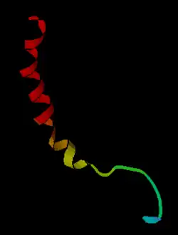
Neuropeptide
Neuropeptides are small proteins produced by neurons that act on G protein-coupled receptors and are responsible for slow-onset, long-lasting modulation of synaptic transmission. Neuropeptides often coexist with each other or with other neurotransmitters in a single neuron. According to their chemical nature, coexisting messengers are localized to different cell compartments: neuropeptides are packaged in large dense core vesicles,[1] whereas low-molecular weight neurotransmitters are stored in small synaptic vesicles.

Neuropeptides conjugated to proteins or other carriers, such as liposomes, may be used for targeting radioisotopes or drugs to cells, specialized endothelia, and normal or neoplastic tissues expressing the corresponding binding sites for diagnostic or therapeutic purposes.
Mechanism and synthesis
Neuropeptides are synthesized from large, inactive precursor proteins called prepropeptides, which are cleaved into several active peptides. Prepropeptides often produce multiple copies of the same peptide or many different peptides.[2] The number of repeats of a peptide sequence often changed throughout evolution and served as a hotbed for genetic variation.
Peptides are synthesized at the soma, entered into the secretory pathway to pass through the rER-Golgi complex, further processed, then packaged into large dense core vesicles for transport down the axon or dendrites.[3][4] The large dense core vesicles are often found in all parts of a neuron, including the soma, dendrites, axonal swellings (varicosities) and nerve endings, whereas the small synaptic vesicles are mainly found in clusters at presynaptic locations.[5][6][1] Release of the large dense core vesicles and the small synaptic vesicles is regulated differently. Neuropeptides are released in a calcium-dependent manner to bind to G protein-coupled receptor(GPCRs)]]. Large dense core vesicles release low volumes of neuropeptide compared to synaptic vesicles and neurotransmitters. Neuropeptides are not immediately reuptaken, degraded or recycled and thus are bioactive for long periods of time.[3]
Peptidergic expression in the brain can be highly selective and specific. In Drosophila larvae for example, eclosion hormone is expressed in just two neurons and SIFamide is expressed in four.[4] In contrast to its selective expression, peptidergic activity can be broad and long-lasting. Neuropeptides are often co-released with other peptides and traditional neurotransmitters. For example, vasoactive intestinal peptide is typically co-released with acetylcholine.[7]
In contrast to its selective expression, peptide action can be broad and diverse. Peptides bind to GPCRs to induce signaling cascades that alter cellular and synaptic activity. There is also tissue-specific processing of neuropeptide precursors. Different tissues have tailored post-translational processing steps which yield structurally and functionally different peptides.[3] Peptides can affect gene expression, local blood flow, synaptogenesis and glial cell morphology.
Receptor targets
Most neuropeptides act on G-protein coupled receptors (GPCRs). Neuropeptide-GPCRs fall into two families: rhodopsin-like and the secretin class.[8] Most peptides activate a single GPCR, while some activate multiple GPCRs (e.g. AstA, AstC, DTK).[9] Peptide-GPCR binding relationships are highly conserved across animals. Aside from conserved structural relationships, some peptide-GPCR functions are also conserved across the animal kingdom. For example, neuropeptide F/neuropeptide Y signaling is structurally and functionally conserved between insects and mammals.[9]
Although peptides mostly target metabotropic receptors, there is some evidence that neuropeptides bind to other receptor targets. Peptide-gated ion channels (FMRFamide-gated sodium channels) have been found in snails and Hydra.[10] Other examples of non-GPCR targets include: insulin-like peptides and tyrosine-kinase receptors in Drosophila and atrial natriuretic peptide and eclosion hormone with membrane-bound guanylyl cyclase receptors in mammals and insects.[11]
Examples
Many populations of neurons have distinctive biochemical phenotypes. For example, in one subpopulation of about 3000 neurons in the arcuate nucleus of the hypothalamus, three anorectic peptides are co-expressed: α-melanocyte-stimulating hormone (α-MSH), galanin-like peptide, and cocaine-and-amphetamine-regulated transcript (CART), and in another subpopulation two orexigenic peptides are co-expressed, neuropeptide Y and agouti-related peptide (AGRP). These are not the only peptides in the arcuate nucleus; β-endorphin, dynorphin, enkephalin, galanin, ghrelin, growth-hormone releasing hormone, neurotensin, neuromedin U, and somatostatin are also expressed in subpopulations of arcuate neurons. These peptides are all released centrally and act on other neurons at specific receptors. The neuropeptide Y neurons also make the classical inhibitory neurotransmitter GABA.
Invertebrates also have many neuropeptides. CCAP has several functions including regulating heart rate, allatostatin and proctolin regulate food intake and growth, bursicon controls tanning of the cuticle and corazonin has a role in cuticle pigmentation and moulting.
Peptide signals play a role in information processing that is different from that of conventional neurotransmitters, and many appear to be particularly associated with specific behaviours. For example, oxytocin and vasopressin have striking and specific effects on social behaviours, including maternal behaviour and pair bonding. The following is a list of neuroactive peptides coexisting with other neurotransmitters. Transmitter names are shown in bold.
Norepinephrine (noradrenaline). In neurons of the A2 cell group in the nucleus of the solitary tract), norepinephrine co-exists with:
- Somatostatin (in the hippocampus)
- Cholecystokinin
- Neuropeptide Y (in the arcuate nucleus)
Epinephrine (adrenaline)
Serotonin (5-HT)
Some neurons make several different peptides. For instance, Vasopressin co-exists with dynorphin and galanin in magnocellular neurons of the supraoptic nucleus and paraventricular nucleus, and with CRF (in parvocellular neurons of the paraventricular nucleus)
Oxytocin in the supraoptic nucleus co-exists with enkephalin, dynorphin, cocaine-and amphetamine regulated transcript (CART) and cholecystokinin.
References
- "Neuronal dense core vesicle". www.uniprot.org. Retrieved 20 December 2020.
- Elphick, Maurice R.; Mirabeau, Olivier; Larhammar, Dan (1 February 2018). "Evolution of neuropeptide signalling systems". Journal of Experimental Biology. 221 (3): jeb151092. doi:10.1242/jeb.151092. ISSN 0022-0949. PMC 5818035. PMID 29440283.
- Mains, Richard E.; Eipper, Betty A. (1999). "The Neuropeptides". Basic Neurochemistry: Molecular, Cellular and Medical Aspects. 6th Edition.
- Nässel, Dick R.; Zandawala, Meet (August 2019). "Recent advances in neuropeptide signaling in Drosophila, from genes to physiology and behavior". Progress in Neurobiology. 179: 101607. doi:10.1016/j.pneurobio.2019.02.003. ISSN 1873-5118. PMID 30905728.
- van den Pol AN (October 2012). "Neuropeptide transmission in brain circuits". Neuron. 76 (1): 98–115. doi:10.1016/j.neuron.2012.09.014. PMC 3918222. PMID 23040809.
- Leng G, Ludwig M (December 2008). "Neurotransmitters and peptides: whispered secrets and public announcements". The Journal of Physiology. 586 (23): 5625–32. doi:10.1113/jphysiol.2008.159103. PMC 2655398. PMID 18845614.
- Dori, I.; Parnavelas, J. G. (1 July 1989). "The cholinergic innervation of the rat cerebral cortex shows two distinct phases in development". Experimental Brain Research. 76 (2): 417–423. doi:10.1007/BF00247899. ISSN 1432-1106. PMID 2767193.
- Brody, Thomas; Cravchik, Anibal (24 July 2000). "Drosophila melanogasterG Protein–Coupled Receptors". Journal of Cell Biology. 150 (2): F83–F88. doi:10.1083/jcb.150.2.F83. ISSN 0021-9525. PMC 2180217. PMID 10908591.
- Nässel, Dick R.; Winther, Åsa M. E. (1 September 2010). "Drosophila neuropeptides in regulation of physiology and behavior". Progress in Neurobiology. 92 (1): 42–104. doi:10.1016/j.pneurobio.2010.04.010. ISSN 0301-0082. PMID 20447440.
- Dürrnagel, Stefan; Kuhn, Anne; Tsiairis, Charisios D.; Williamson, Michael; Kalbacher, Hubert; Grimmelikhuijzen, Cornelis J. P.; Holstein, Thomas W.; Gründer, Stefan (16 April 2010). "Three Homologous Subunits Form a High Affinity Peptide-gated Ion Channel in Hydra". Journal of Biological Chemistry. 285 (16): 11958–11965. doi:10.1074/jbc.M109.059998. ISSN 0021-9258. PMC 2852933. PMID 20159980.
- Chang, Jer-Cherng; Yang, Ruey-Bing; Adams, Michael E.; Lu, Kuang-Hui (11 August 2009). "Receptor guanylyl cyclases in Inka cells targeted by eclosion hormone". Proceedings of the National Academy of Sciences. 106 (32): 13371–13376. Bibcode:2009PNAS..10613371C. doi:10.1073/pnas.0812593106. ISSN 0027-8424. PMC 2726410. PMID 19666575.
| Wikimedia Commons has media related to Neuropeptides. |
External links
| Look up neuropeptide in Wiktionary, the free dictionary. |
- Neuropeptides Journal
- Neuropeptides reference website (a comprehensive neuropeptide database)
- Neuropeptides eBook series
- Neuropeptide chapter in the C. elegans Wormbook excellent, and very accessible, discussion of neuropeptide biology in C. elegans