Squalene monooxygenase
Squalene monooxygenase (also called squalene epoxidase) is an enzyme that uses NADPH and molecular oxygen to oxidize squalene to 2,3-oxidosqualene (squalene epoxide). Squalene epoxidase catalyzes the first oxygenation step in sterol biosynthesis and is thought to be one of the rate-limiting enzymes in this pathway.[5] In humans, squalene epoxidase is encoded by the SQLE gene.[6] Several eukaryote genomes lack a squalene monooxygenase encoding gene, but instead encode an alternative squalene epoxidase that catalyzes the oxidation of squalene.[7]
| Squalene epoxidase | |||||||||
|---|---|---|---|---|---|---|---|---|---|
 Chemical reaction catalyzed by squalene epoxidase. | |||||||||
| Identifiers | |||||||||
| EC number | 1.14.13.132 | ||||||||
| CAS number | 9029-62-3 | ||||||||
| Databases | |||||||||
| IntEnz | IntEnz view | ||||||||
| BRENDA | BRENDA entry | ||||||||
| ExPASy | NiceZyme view | ||||||||
| KEGG | KEGG entry | ||||||||
| MetaCyc | metabolic pathway | ||||||||
| PRIAM | profile | ||||||||
| PDB structures | RCSB PDB PDBe PDBsum | ||||||||
| Gene Ontology | AmiGO / QuickGO | ||||||||
| |||||||||
Mechanism
The canonical squalene monooxygenase is a flavoprotein monooxygenase. Flavoprotein monooxygenase form flavin hydroperoxides at the enzyme active site, which then transfer the terminal oxygen atom of the hydroperoxide to the substrate. Squalene monooxygenase differs from other flavin monooxygenases in that the oxygen is inserted as an epoxide rather than as a hydroxyl group. Squalene monooxygenase contains a loosely bound FAD flavin and obtains electrons from NADPH-cytochrome P450 reductase, rather than binding the nicotinamide cofactor NADPH directly. The alternative squalene epoxidase belongs to the fatty acid hydroxylase superfamily and obtains electrons from cytochrome b5.[7]
Inhibitors
Inhibitors of squalene epoxidase have found application mainly as antifungal drugs:[8]
Since squalene epoxidase is on the biosynthetic pathway leading to cholesterol, inhibitors of this enzyme may also find application in treatment of hypercholesterolemia.[10]
Localization
In yeast Saccharomyces cerevisiae, squalene epoxidase is localized to both the endoplasmic reticulum and lipid droplets. Only the ER localized protein is active.
Additional products
Squalene epoxidase also catalyzes the formation of diepoxysqualene (DOS). DOS is converted to 24(S),25-epoxylanosterol by lanosterol synthase.
Model organisms
Model organisms have been used in the study of SQLE function. A conditional knockout mouse line called Sqletm1a(EUCOMM)Wtsi was generated at the Wellcome Trust Sanger Institute.[11] Male and female animals underwent a standardized phenotypic screen[12] to determine the effects of deletion.[13][14][15][16] Additional screens performed: - In-depth immunological phenotyping[17]
| Characteristic | Phenotype |
|---|---|
| All data available at.[12][17] | |
| Peripheral blood leukocytes 6 Weeks | Normal |
| Insulin | Normal |
| Haematology 6 Weeks | Normal |
| Homozygous viability at P14 | Abnormal |
| Recessive lethal study | Abnormal |
| Body weight | Normal |
| Neurological assessment | Normal |
| Grip strength | Normal |
| Dysmorphology | Normal |
| Indirect calorimetry | Normal |
| Glucose tolerance test | Normal |
| Auditory brainstem response | Normal |
| DEXA | Normal |
| Radiography | Normal |
| Eye morphology | Normal |
| Clinical chemistry | Normal |
| Haematology 16 Weeks | Normal |
| Peripheral blood leukocytes 16 Weeks | Normal |
| Heart weight | Normal |
| Salmonella infection | Normal |
| Cytotoxic T Cell Function | Normal |
| Spleen Immunophenotyping | Normal |
| Mesenteric Lymph Node Immunophenotyping | Normal |
| Bone Marrow Immunophenotyping | Normal |
| Epidermal Immune Composition | Normal |
See also
References
- GRCh38: Ensembl release 89: ENSG00000104549 - Ensembl, May 2017
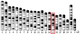
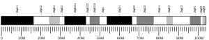
- GRCm38: Ensembl release 89: ENSMUSG00000022351 - Ensembl, May 2017
- "Human PubMed Reference:". National Center for Biotechnology Information, U.S. National Library of Medicine.
- "Mouse PubMed Reference:". National Center for Biotechnology Information, U.S. National Library of Medicine.
- "Entrez Gene: SQLE squalene epoxidase".
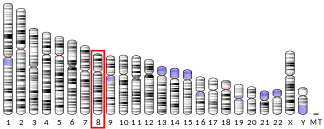
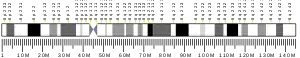
- Nagai M, Sakakibara J, Wakui K, Fukushima Y, Igarashi S, Tsuji S, Arakawa M, Ono T (Aug 1997). "Localization of the squalene epoxidase gene (SQLE) to human chromosome region 8q24.1". Genomics. 44 (1): 141–3. doi:10.1006/geno.1997.4825. PMID 9286711.
- Pollier J, Vancaester E, Kuzhiumparambil U, Vickers CE, Vandepoele K, Goossens A, Fabris M (2019). "A widespread alternative squalene epoxidase participates in eukaryote steroid biosynthesis". Nature Microbiology. 4 (2): 226–233. doi:10.1038/s41564-018-0305-5. PMID 30478288. S2CID 53726187.
- Favre B, Ryder NS (Feb 1996). "Characterization of squalene epoxidase activity from the dermatophyte Trichophyton rubrum and its inhibition by terbinafine and other antimycotic agents". Antimicrobial Agents and Chemotherapy. 40 (2): 443–7. doi:10.1128/AAC.40.2.443. PMC 163131. PMID 8834895.
- Ryder NS (Feb 1992). "Terbinafine: mode of action and properties of the squalene epoxidase inhibition". The British Journal of Dermatology. 126 Suppl 39: 2–7. doi:10.1111/j.1365-2133.1992.tb00001.x. PMID 1543672. S2CID 19780957.
- Chugh A, Ray A, Gupta JB (Jan 2003). "Squalene epoxidase as hypocholesterolemic drug target revisited". Progress in Lipid Research. 42 (1): 37–50. doi:10.1016/S0163-7827(02)00029-2. PMID 12467639.
- Gerdin AK (2010). "The Sanger Mouse Genetics Programme: high throughput characterisation of knockout mice". Acta Ophthalmologica. 88: 925–7. doi:10.1111/j.1755-3768.2010.4142.x. S2CID 85911512.
- "International Mouse Phenotyping Consortium".
- Skarnes WC, Rosen B, West AP, Koutsourakis M, Bushell W, Iyer V, Mujica AO, Thomas M, Harrow J, Cox T, Jackson D, Severin J, Biggs P, Fu J, Nefedov M, de Jong PJ, Stewart AF, Bradley A (Jun 2011). "A conditional knockout resource for the genome-wide study of mouse gene function". Nature. 474 (7351): 337–42. doi:10.1038/nature10163. PMC 3572410. PMID 21677750.
- Dolgin E (Jun 2011). "Mouse library set to be knockout". Nature. 474 (7351): 262–3. doi:10.1038/474262a. PMID 21677718.
- Collins FS, Rossant J, Wurst W (Jan 2007). "A mouse for all reasons". Cell. 128 (1): 9–13. doi:10.1016/j.cell.2006.12.018. PMID 17218247. S2CID 18872015.
- White JK, Gerdin AK, Karp NA, Ryder E, Buljan M, Bussell JN, Salisbury J, Clare S, Ingham NJ, Podrini C, Houghton R, Estabel J, Bottomley JR, Melvin DG, Sunter D, Adams NC, Tannahill D, Logan DW, Macarthur DG, Flint J, Mahajan VB, Tsang SH, Smyth I, Watt FM, Skarnes WC, Dougan G, Adams DJ, Ramirez-Solis R, Bradley A, Steel KP (Jul 2013). "Genome-wide generation and systematic phenotyping of knockout mice reveals new roles for many genes". Cell. 154 (2): 452–64. doi:10.1016/j.cell.2013.06.022. PMC 3717207. PMID 23870131.
- "Infection and Immunity Immunophenotyping (3i) Consortium".
Further reading
- Ma J, Dempsey AA, Stamatiou D, Marshall KW, Liew CC (Mar 2007). "Identifying leukocyte gene expression patterns associated with plasma lipid levels in human subjects". Atherosclerosis. 191 (1): 63–72. doi:10.1016/j.atherosclerosis.2006.05.032. PMID 16806233.
- Laden BP, Tang Y, Porter TD (Feb 2000). "Cloning, heterologous expression, and enzymological characterization of human squalene monooxygenase". Archives of Biochemistry and Biophysics. 374 (2): 381–8. doi:10.1006/abbi.1999.1629. PMID 10666321.
- Helms MW, Kemming D, Pospisil H, Vogt U, Buerger H, Korsching E, Liedtke C, Schlotter CM, Wang A, Chan SY, Brandt BH (Sep 2008). "Squalene epoxidase, located on chromosome 8q24.1, is upregulated in 8q+ breast cancer and indicates poor clinical outcome in stage I and II disease". British Journal of Cancer. 99 (5): 774–80. doi:10.1038/sj.bjc.6604556. PMC 2528137. PMID 18728668.
- Nagai M, Sakakibara J, Nakamura Y, Gejyo F, Ono T (Jul 2002). "SREBP-2 and NF-Y are involved in the transcriptional regulation of squalene epoxidase". Biochemical and Biophysical Research Communications. 295 (1): 74–80. doi:10.1016/S0006-291X(02)00623-X. PMID 12083769.
- Liu Y, Sun W, Zhang K, Zheng H, Ma Y, Lin D, Zhang X, Feng L, Lei W, Zhang Z, Guo S, Han N, Tong W, Feng X, Gao Y, Cheng S (Jun 2007). "Identification of genes differentially expressed in human primary lung squamous cell carcinoma". Lung Cancer. 56 (3): 307–17. doi:10.1016/j.lungcan.2007.01.016. PMID 17316888.
- Suzuki Y, Yoshitomo-Nakagawa K, Maruyama K, Suyama A, Sugano S (Oct 1997). "Construction and characterization of a full length-enriched and a 5'-end-enriched cDNA library". Gene. 200 (1–2): 149–56. doi:10.1016/S0378-1119(97)00411-3. PMID 9373149.
- Lu Y, Dollé ME, Imholz S, van 't Slot R, Verschuren WM, Wijmenga C, Feskens EJ, Boer JM (Dec 2008). "Multiple genetic variants along candidate pathways influence plasma high-density lipoprotein cholesterol concentrations". Journal of Lipid Research. 49 (12): 2582–9. doi:10.1194/jlr.M800232-JLR200. PMID 18660489.
- Mehrle A, Rosenfelder H, Schupp I, del Val C, Arlt D, Hahne F, Bechtel S, Simpson J, Hofmann O, Hide W, Glatting KH, Huber W, Pepperkok R, Poustka A, Wiemann S (Jan 2006). "The LIFEdb database in 2006". Nucleic Acids Research. 34 (Database issue): D415-8. doi:10.1093/nar/gkj139. PMC 1347501. PMID 16381901.
- Hartley JL, Temple GF, Brasch MA (Nov 2000). "DNA cloning using in vitro site-specific recombination". Genome Research. 10 (11): 1788–95. doi:10.1101/gr.143000. PMC 310948. PMID 11076863.
- Maruyama K, Sugano S (Jan 1994). "Oligo-capping: a simple method to replace the cap structure of eukaryotic mRNAs with oligoribonucleotides". Gene. 138 (1–2): 171–4. doi:10.1016/0378-1119(94)90802-8. PMID 8125298.
- Nakamura Y, Sakakibara J, Izumi T, Shibata A, Ono T (Apr 1996). "Transcriptional regulation of squalene epoxidase by sterols and inhibitors in HeLa cells". The Journal of Biological Chemistry. 271 (14): 8053–6. doi:10.1074/jbc.271.14.8053. PMID 8626488.
- Nagai M, Sakakibara J, Wakui K, Fukushima Y, Igarashi S, Tsuji S, Arakawa M, Ono T (Aug 1997). "Localization of the squalene epoxidase gene (SQLE) to human chromosome region 8q24.1". Genomics. 44 (1): 141–3. doi:10.1006/geno.1997.4825. PMID 9286711.
- Wiemann S, Arlt D, Huber W, Wellenreuther R, Schleeger S, Mehrle A, Bechtel S, Sauermann M, Korf U, Pepperkok R, Sültmann H, Poustka A (Oct 2004). "From ORFeome to biology: a functional genomics pipeline". Genome Research. 14 (10B): 2136–44. doi:10.1101/gr.2576704. PMC 528930. PMID 15489336.
External links
- Squalene+monooxygenase at the US National Library of Medicine Medical Subject Headings (MeSH)
This article incorporates text from the United States National Library of Medicine, which is in the public domain.