Biomineralization
Biomineralization, or biomineralisation is the process by which living organisms produce minerals,[lower-alpha 1] often to harden or stiffen existing tissues. Such tissues are called mineralized tissues. It is an extremely widespread phenomenon; all six taxonomic kingdoms contain members that are able to form minerals, and over 60 different minerals have been identified in organisms.[3][4][5] Examples include silicates in algae and diatoms, carbonates in invertebrates, and calcium phosphates and carbonates in vertebrates. These minerals often form structural features such as sea shells and the bone in mammals and birds. Organisms have been producing mineralised skeletons for the past 550 million years. Ca carbonates and Ca phosphates are usually crystalline, but silica organisms (sponges, diatoms...) are always non crystalline minerals. Other examples include copper, iron and gold deposits involving bacteria. Biologically-formed minerals often have special uses such as magnetic sensors in magnetotactic bacteria (Fe3O4), gravity sensing devices (CaCO3, CaSO4, BaSO4) and iron storage and mobilization (Fe2O3•H2O in the protein ferritin).
| Part of a series related to |
| Biomineralization |
|---|
 |
In terms of taxonomic distribution, the most common biominerals are the phosphate and carbonate salts of calcium that are used in conjunction with organic polymers such as collagen and chitin to give structural support to bones and shells.[6] The structures of these biocomposite materials are highly controlled from the nanometer to the macroscopic level, resulting in complex architectures that provide multifunctional properties. Because this range of control over mineral growth is desirable for materials engineering applications, there is significant interest in understanding and elucidating the mechanisms of biologically controlled biomineralization.[7][8]
Types of mineralization
Mineralization can be subdivided into different categories depending on the following: the organisms or processes that create chemical conditions necessary for mineral formation, the origin of the substrate at the site of mineral precipitation, and the degree of control that the substrate has on crystal morphology, composition, and growth.[9] These subcategories include: biomineralization, organomineralization, and inorganic mineralization, which can be subdivided further. However, usage of these terms vary widely in scientific literature because there are no standardized definitions. The following definitions are based largely on a paper written by Dupraz et al. (2009), which provided a framework for differentiating these terms.
Biomineralization
Biomineralization, biologically-controlled mineralization, occurs when crystal morphology, growth, composition, and location is completely controlled by the cellular processes of a specific organism. Examples include the shells of invertebrates, such as molluscs and brachiopods. Additionally, mineralization of collagen provides the crucial compressive strength for the bones, cartilage, and teeth of vertebrates.[10]
Organomineralization
This type of mineralization includes both biologically-induced mineralization and biologically-influenced mineralization.
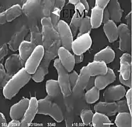
- Biologically-induced mineralization occurs when the metabolic activity of microbes (e.g. bacteria) produces chemical conditions favorable for mineral formation. The substrate for mineral growth is the organic matrix, secreted by the microbial community, and affects crystal morphology and composition. Examples of this type of mineralization include calcareous or siliceous stromatolites and other microbial mats. A more specific type of biologically-induced mineralization, remote calcification or remote mineralization, takes place when calcifying microbes occupy a shell secreting organism and alter the chemical environment surrounding the area of shell formation. The result is mineral formation not strongly controlled by the cellular processes of the metazoan host (i.e. remote mineralization), and may lead to unique or unusual crystal morphologies.[11]
- Biologically-influenced mineralization takes place when chemical conditions surrounding the site of mineral formation are influenced by abiotic processes (e.g. evaporation or degassing). However, the organic matrix (secreted by microorganisms) is responsible for crystal morphology and composition. Examples include micro- to nano-meter scale crystals of various morphologies.
Inorganic mineralization
Inorganic mineralization is a completely abiotic process. Chemical conditions necessary for mineral formation develop via environmental processes, such as evaporation or degassing. Furthermore, the substrate for mineral deposition is abiotic (i.e. contains no organic compounds) and there is no control on crystal morphology or composition. Examples of this type of mineralization include cave formations, such as stalagmites and stalactites.
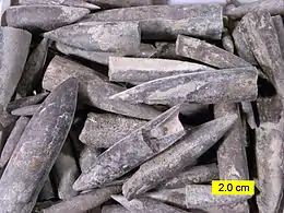
Biological mineralization can also take place as a result of fossilization. See also calcification.
Bone mineralization occurs in human body by cells called osteoblasts.
Biological roles
Among metazoans, biominerals composed of calcium carbonate, calcium phosphate or silica perform a variety of roles such as support, defense and feeding.[12] It is less clear what purpose biominerals serve in bacteria. One hypothesis is that cells create them to avoid entombment by their own metabolic byproducts. Iron oxide particles may also enhance their metabolism.[13]
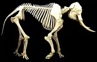 Vertebrate animals have internal endoskeletons which achieve rigidity by binding calcium phosphate into hydroxylapatite
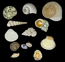
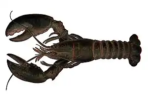 Many invertebrate animals have external exoskeletons or shells, which achieve rigidity by a variety of mineralisations
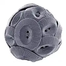
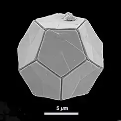 Many protists, like this coccolithophore, have protective mineralised shells
Biology
If present on a super-cellular scale, biominerals are usually deposited by a dedicated organ, which is often defined very early in the embryological development. This organ will contain an organic matrix that facilitates and directs the deposition of crystals.[12] The matrix may be collagen, as in deuterostomes,[12] or based on chitin or other polysaccharides, as in molluscs.[14]
Shell formation in molluscs
The mollusc shell is a biogenic composite material that has been the subject of much interest in materials science because of its unusual properties and its model character for biomineralization. Molluscan shells consist of 95–99% calcium carbonate by weight, while an organic component makes up the remaining 1–5%. The resulting composite has a fracture toughness ≈3000 times greater than that of the crystals themselves.[15] In the biomineralization of the mollusc shell, specialized proteins are responsible for directing crystal nucleation, phase, morphology, and growths dynamics and ultimately give the shell its remarkable mechanical strength. The application of biomimetic principles elucidated from mollusc shell assembly and structure may help in fabricating new composite materials with enhanced optical, electronic, or structural properties. The most described arrangement in mollusc shells is the nacre - prismatic shells, known in large shells as Pinna or the pearl oyster (Pinctada). Not only the structure of the layers differ, but their mineralogy and chemical composition also differ. Both contain organic components (proteins, sugars and lipids) and the organic components are characteristic of the layer, and of the species.[5] The structures and arrangements of mollusc shells are diverse, but they share some features: the main part of the shell is a crystalline Ca carbonate (aragonite, calcite), despite some amorphous Ca carbonate occurs; and despite they react as crystals, they never show angles and facets.[16] The examination of the inner structure of the prismatic units, nacreous tablets, foliated laths... shows irregular rounded granules.
Mineral production and degradation in fungi
Fungi are a diverse group of organisms that belong to the eukaryotic domain. Studies of their significant roles in geological processes, "geomycology", has shown that fungi are involved with biomineralization, biodegradation, and metal-fungal interactions.[17]
In studying fungi's roles in biomineralization, it has been found that fungi deposit minerals with the help of an organic matrix, such as a protein, that provides a nucleation site for the growth of biominerals.[18] Fungal growth may produce a copper-containing mineral precipitate, such as copper carbonate produced from a mixture of (NH4)2CO3 and CuCl2.[18] The production of the copper carbonate is produced in the presence of proteins made and secreted by the fungi.[18] These fungal proteins that are found extracellularly aid in the size and morphology of the carbonate minerals precipitated by the fungi.[18]
In addition to precipitating carbonate minerals, fungi can also precipitate uranium-containing phosphate biominerals in the presence of organic phosphorus that acts a substrate for the process.[19] The fungi produce a hyphal matrix, also known as mycelium, that localizes and accumulates the uranium minerals that have been precipitated.[19] Although uranium is often deemed as toxic towards living organisms, certain fungi such as Aspergillus niger and Paecilomyces javanicus can tolerate it.
Though minerals can be produced by fungi, they can also be degraded; mainly by oxalic-acid producing strains of fungi.[20] Oxalic acid production is increased in the presence of glucose for three organic acid producing fungi – Aspergillus niger, Serpula himantioides, and Trametes versicolor.[20] These fungi have been found to corrode apatite and galena minerals.[20] Degradation of minerals by fungi is carried out through a process known as neogenesis.[21] The order of most to least oxalic acid secreted by the fungi studied are Aspergillus niger, followed by Serpula himantioides, and finally Trametes versicolor.[20] These capabilities of certain groups of fungi have a major impact on corrosion, a costly problem for many industries and the economy.
Chemistry
The majority of biominerals fall into three distinct mineral classes: carbonates, silicates and phosphates.[22]
Silicates
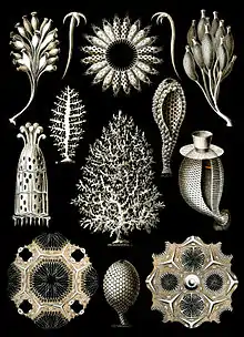
Silicates are particularly common in marine biominerals, where diatoms and radiolaria form frustules from hydrated amorphous silica (opal).
Carbonates
The major carbonates are CaCO3. The most common polymorphs in biomineralization are calcite (e.g. foraminifera, coccolithophores) and aragonite (e.g. corals), although metastable vaterite and amorphous calcium carbonate can also be important, either structurally [23][24] or as intermediate phases in biomineralization.[25][26] Some biominerals include a mixture of these phases in distinct, organised structural components (e.g. bivalve shells). Carbonates are particularly prevalent in marine environments, but also present in fresh water and terrestrial organisms.
Phosphates
The most common phosphate is hydroxyapatite, a calcium phosphate (Ca10(PO4)6(OH)2) which is a primary constituent of bone, teeth, and fish scales.[27]
Other minerals
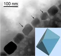
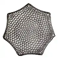
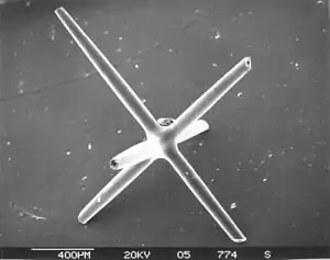
Beyond these main three categories, there are a number of less common types of biominerals, usually resulting from a need for specific physical properties or the organism inhabiting an unusual environment. For example, teeth that are primarily used for scraping hard substrates may be reinforced with particularly tough minerals, such as the iron minerals magnetite in chiton,[28] or goethite in limpets.[29] Gastropod molluscs living close to hydrothermal vents reinforce their carbonate shells with the iron-sulphur minerals pyrite and greigite.[30] Magnetotactic bacteria also employ magnetic iron minerals magnetite and greigite to produce magnetosomes to aid orientation and distribution in the sediments. Planktic acantharea (radiolaria) form strontium sulphate (celestine) spicules.
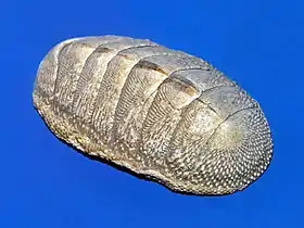 Chiton have aragonite shells and teeth coated with magnetite
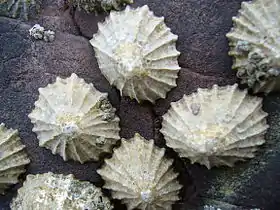 Limpets have carbonate shells and teeth reinforced with goethite
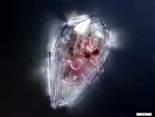
.jpg.webp) Acantharian radiolarians have strontium sulfate crystal shells
Acantharian radiolarians have strontium sulfate crystal shells
Diversity
_mitra_Ehrenberg_-_160x.jpg.webp)
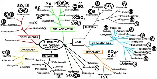
In nature, there is a wide array of biominerals, ranging from iron oxide to strontium sulfate,[37] with calcareous biominerals being particularly notable.[39][40] However, the most taxonomically widespread biomineral is silica (SiO2·nH2O), being present in all eukaryotic supergroups.[41] Notwithstanding, the degree of silicification can vary even between closely related taxa, from being found in composite structures with other biominerals (e.g., limpet teeth;[42] to forming minor structures (e.g., ciliate granules;[43] or being a major structural constituent of the organism.[44] The most extreme degree of silicification is evident in the diatoms, where almost all species have an obligate requirement for silicon to complete cell wall formation and cell division.[45][46] Biogeochemically and ecologically, diatoms are the most important silicifiers in modern marine ecosystems, with radiolarians (polycystine and phaeodarian rhizarians), silicoflagellates (dictyochophyte and chrysophyte stramenopiles), and sponges with prominent roles as well. In contrast, the major silicifiers in terrestrial ecosystems are the land plants (embryophytes), with other silicifying groups (e.g., testate amoebae) having a minor role.[32]
Broadly, biomineralized structures evolve and diversify when the energetic cost of biomineral production is less than the expense of producing an equivalent organic structure.[47][48][49] The energetic costs of forming a silica structure from silicic acid are much less than forming the same volume from an organic structure (~20x less than lignin or 10x less than polysaccharides like cellulose).[50] Based on a structural model of biogenic silica,[51] Lobel et al. (1996) identified by biochemical modeling a low-energy reaction pathway for nucleation and growth of silica.[52] The combination of organic and inorganic components within biomineralized structures often results in enhanced properties compared to exclusively organic or inorganic materials. With respect to biogenic silica, this can result in the production of much stronger structures, such as siliceous diatom frustules having the highest strength per unit density of any known biological material,[53][54] or sponge spicules being many times more flexible than an equivalent structure made of pure silica.[55][56] As a result, biogenic silica structures are utilized for support,[57] feeding,[58] predation defense [59][60][61] and environmental protection as a component of cyst walls.[44] Biogenic silica also has useful optical properties for light transmission and modulation in organisms as diverse as plants,[62] diatoms,[63][64][65] sponges,[66] and molluscs.[67] There is also evidence that silicification is used as a detoxification response in snails [68] and plants,[69] biosilica has even been suggested to play a role as a pH buffer for the enzymatic activity of carbonic anhydrase, aiding the acquisition of inorganic carbon for photosynthesis.[70][32]
There are questions which have yet to be resolved, such as why do some organisms biomineralize while others do not, and why is there such a diversity of biominerals besides silicon when silicon is so abundant, comprising 28% of the Earth's crust.[32] The answer to these questions lies in the evolutionary interplay between biomineralization and geochemistry, and in the competitive interactions that have arisen from these dynamics. Fundamentally whether an organism produces silica or not involves evolutionary trade-offs and competition between silicifiers themselves, and with non-silicifying organisms (both those which utilize other biominerals, and non-mineralizing groups). Mathematical models and controlled experiments of resource competition in phytoplankton have demonstrated the rise to dominance of different algal species based on nutrient backgrounds in defined media. These have been part of fundamental studies in ecology.[71] [72] However, the vast diversity of organisms that thrive in a complex array of biotic and abiotic interactions in oceanic ecosystems are a challenge to such minimal models and experimental designs, whose parameterization and possible combinations, respectively, limit the interpretations that can be built on them.[32]
Evolution
The first evidence of biomineralization dates to some 750 million years ago,[73][74] and sponge-grade organisms may have formed calcite skeletons 630 million years ago.[75] But in most lineages, biomineralization first occurred in the Cambrian or Ordovician periods.[76] Organisms used whichever form of calcium carbonate was more stable in the water column at the point in time when they became biomineralized,[77] and stuck with that form for the remainder of their biological history[78] (but see [79] for a more detailed analysis). The stability is dependent on the Ca/Mg ratio of seawater, which is thought to be controlled primarily by the rate of sea floor spreading, although atmospheric CO
2 levels may also play a role.[77]
Biomineralization evolved multiple times, independently,[80] and most animal lineages first expressed biomineralized components in the Cambrian period.[81] Many of the same processes are used in unrelated lineages, which suggests that biomineralization machinery was assembled from pre-existing "off-the-shelf" components already used for other purposes in the organism.[22] Although the biomachinery facilitating biomineralization is complex – involving signalling transmitters, inhibitors, and transcription factors – many elements of this 'toolkit' are shared between phyla as diverse as corals, molluscs, and vertebrates.[82] The shared components tend to perform quite fundamental tasks, such as designating that cells will be used to create the minerals, whereas genes controlling more finely tuned aspects that occur later in the biomineralization process – such as the precise alignment and structure of the crystals produced – tend to be uniquely evolved in different lineages.[12][83] This suggests that Precambrian organisms were employing the same elements, albeit for a different purpose — perhaps to avoid the inadvertent precipitation of calcium carbonate from the supersaturated Proterozoic oceans.[82] Forms of mucus that are involved in inducing mineralization in most metazoan lineages appear to have performed such an anticalcifatory function in the ancestral state.[84] Further, certain proteins that would originally have been involved in maintaining calcium concentrations within cells[85] are homologous to all metazoans, and appear to have been co-opted into biomineralization after the divergence of the metazoan lineages.[86] The galaxins are one probable example of a gene being co-opted from a different ancestral purpose into controlling biomineralization, in this case being 'switched' to this purpose in the Triassic scleractinian corals; the role performed appears to be functionally identical to the unrelated pearlin gene in molluscs.[87] Carbonic anhydrase serves a role in mineralization in sponges, as well as metazoans, implying an ancestral role.[88] Far from being a rare trait that evolved a few times and remained stagnant, biomineralization pathways in fact evolved many times and are still evolving rapidly today; even within a single genus it is possible to detect great variation within a single gene family.[83]
The homology of biomineralization pathways is underlined by a remarkable experiment whereby the nacreous layer of a molluscan shell was implanted into a human tooth, and rather than experiencing an immune response, the molluscan nacre was incorporated into the host bone matrix. This points to the exaptation of an original biomineralization pathway.
The most ancient example of biomineralization, dating back 2 billion years, is the deposition of magnetite, which is observed in some bacteria, as well as the teeth of chitons and the brains of vertebrates; it is possible that this pathway, which performed a magnetosensory role in the common ancestor of all bilaterians, was duplicated and modified in the Cambrian to form the basis for calcium-based biomineralization pathways.[89] Iron is stored in close proximity to magnetite-coated chiton teeth, so that the teeth can be renewed as they wear. Not only is there a marked similarity between the magnetite deposition process and enamel deposition in vertebrates but some vertebrates even have comparable iron storage facilities near their teeth.[90]

| Type of mineralization | Examples of organisms |
|---|---|
| Calcium carbonate (calcite or aragonite) | |
| Silica |
|
| Apatite (phosphate carbonate) |
|
Potential applications
Most traditional approaches to synthesis of nanoscale materials are energy inefficient, requiring stringent conditions (e.g., high temperature, pressure or pH) and often produce toxic byproducts. Furthermore, the quantities produced are small, and the resultant material is usually irreproducible because of the difficulties in controlling agglomeration.[91] In contrast, materials produced by organisms have properties that usually surpass those of analogous synthetically manufactured materials with similar phase composition. Biological materials are assembled in aqueous environments under mild conditions by using macromolecules. Organic macromolecules collect and transport raw materials and assemble these substrates and into short- and long-range ordered composites with consistency and uniformity. The aim of biomimetics is to mimic the natural way of producing minerals such as apatites. Many man-made crystals require elevated temperatures and strong chemical solutions, whereas the organisms have long been able to lay down elaborate mineral structures at ambient temperatures. Often, the mineral phases are not pure but are made as composites that entail an organic part, often protein, which takes part in and controls the biomineralisation. These composites are often not only as hard as the pure mineral but also tougher, as the micro-environment controls biomineralisation.
Architecture
of bacterial biofilms
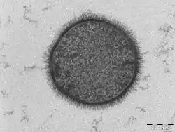
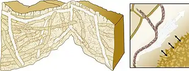
One biological system that might be of key importance in future development of architecture is the bacterial biofilm. The term biofilm refers to complex heterogeneous structures comprising different populations of microorganisms that attach and form a community on an inert (e.g. rocks, glass, plastic) or organic (e.g. skin, cuticle, mucosa) surfaces.[94] The properties of the surface, such as charge, hydrophobicity and roughness, determine initial bacterial attachment.[95] A common principle of all biofilms is the production of extracellular matrix (ECM) composed of different organic substances, such as extracellular proteins, exopolysaccharides and nucleic acids.[96] While the ability to generate ECM appears to be a common feature of multicellular bacterial communities, the means by which these matrices are constructed and function are diverse.[96][97][98][93]
Bacterially induced calcium carbonate precipitation can be used to produce "self‐healing" concrete. Bacillus megaterium spores and suitable dried nutrients are mixed and applied to steel‐reinforced concrete. When the concrete cracks, water ingress dissolves the nutrients and the bacteria germinate triggering calcium carbonate precipitation, resealing the crack and protecting the steel reinforcement from corrosion.[99] This process can also be used to manufacture new hard materials, such as bio‐cement.[100][93]
However the full potential of bacteria‐driven biomineralization is yet to be realized, as it is currently used as a passive filling rather than as a smart designable material. A future challenge is to develop ways to control the timing and the location of mineral formation, as well as the physical properties of the mineral itself, by environmental input. Bacillus subtilis has already been shown to respond to its environment, by changing the production of its ECM. It uses the polymers produced by single cells during biofilm formation as a physical cue to coordinate ECM production by the bacterial community.[101][102][93]
Uranium contaminants in groundwater

Biomineralization may be used to remediate groundwater contaminated with uranium.[103] The biomineralization of uranium primarily involves the precipitation of uranium phosphate minerals associated with the release of phosphate by microorganisms. Negatively charged ligands at the surface of the cells attract the positively charged uranyl ion (UO22+). If the concentrations of phosphate and UO22+ are sufficiently high, minerals such as autunite (Ca(UO2)2(PO4)2•10-12H2O) or polycrystalline HUO2PO4 may form thus reducing the mobility of UO22+. Compared to the direct addition of inorganic phosphate to contaminated groundwater, biomineralization has the advantage that the ligands produced by microbes will target uranium compounds more specifically rather than react actively with all aqueous metals. Stimulating bacterial phosphatase activity to liberate phosphate under controlled conditions limits the rate of bacterial hydrolysis of organophosphate and the release of phosphate to the system, thus avoiding clogging of the injection location with metal phosphate minerals.[103] The high concentration of ligands near the cell surface also provides nucleation foci for precipitation, which leads to higher efficiency than chemical precipitation.[104]
The biogenic mineral controversy
The geological definition of mineral normally excludes compounds that occur only in living beings. However some minerals are often biogenic (such as calcite) or are organic compounds in the sense of chemistry (such as mellite). Moreover, living beings often synthesize inorganic minerals (such as hydroxylapatite) that also occur in rocks.
The International Mineralogical Association (IMA) is the generally recognized standard body for the definition and nomenclature of mineral species. As of December 2020, the IMA recognizes 5,650 official mineral species[105] out of 5,862 proposed or traditional ones.[106]
A topic of contention among geologists and mineralogists has been the IMA's decision to exclude biogenic crystalline substances. For example, Lowenstam (1981) stated that "organisms are capable of forming a diverse array of minerals, some of which cannot be formed inorganically in the biosphere."[107]
Skinner (2005) views all solids as potential minerals and includes biominerals in the mineral kingdom, which are those that are created by the metabolic activities of organisms. Skinner expanded the previous definition of a mineral to classify "element or compound, amorphous or crystalline, formed through biogeochemical processes," as a mineral.[108]
Recent advances in high-resolution genetics and X-ray absorption spectroscopy are providing revelations on the biogeochemical relations between microorganisms and minerals that may shed new light on this question.[109][108] For example, the IMA-commissioned "Working Group on Environmental Mineralogy and Geochemistry " deals with minerals in the hydrosphere, atmosphere, and biosphere.[110] The group's scope includes mineral-forming microorganisms, which exist on nearly every rock, soil, and particle surface spanning the globe to depths of at least 1600 metres below the sea floor and 70 kilometres into the stratosphere (possibly entering the mesosphere).[111][112][113]
Biogeochemical cycles have contributed to the formation of minerals for billions of years. Microorganisms can precipitate metals from solution, contributing to the formation of ore deposits. They can also catalyze the dissolution of minerals.[114][115][116]
Prior to the International Mineralogical Association's listing, over 60 biominerals had been discovered, named, and published.[117] These minerals (a sub-set tabulated in Lowenstam (1981)[107]) are considered minerals proper according to Skinner's (2005) definition.[108] These biominerals are not listed in the International Mineral Association official list of mineral names,[118] however, many of these biomineral representatives are distributed amongst the 78 mineral classes listed in the Dana classification scheme.[108]
Skinner's (2005) definition of a mineral takes this matter into account by stating that a mineral can be crystalline or amorphous.[108] Although biominerals are not the most common form of minerals,[119] they help to define the limits of what constitutes a mineral proper. Nickel's (1995) formal definition explicitly mentioned crystallinity as a key to defining a substance as a mineral.[109] A 2011 article defined icosahedrite, an aluminium-iron-copper alloy as mineral; named for its unique natural icosahedral symmetry, it is a quasicrystal. Unlike a true crystal, quasicrystals are ordered but not periodic.[120][121]
List of minerals
Examples of biogenic minerals include:[122]
- Apatite in bones and teeth.
- Aragonite, calcite, fluorite in vestibular systems (part of the inner ear) of vertebrates.
- Aragonite and calcite in travertine and biogenic silica (siliceous sinter, opal) deposited through algal action.
- Hydroxylapatite formed by mitochondria.
- Magnetite and greigite formed by magnetotactic bacteria.
- Pyrite and marcasite in sedimentary rocks deposited by sulfate-reducing bacteria.
- Quartz and diamonds formed from bacterial action on fossil fuels (gas, oil, coal).
- Goethite found as filaments in limpet teeth.
Astrobiology
It has been suggested that biominerals could be important indicators of extraterrestrial life and thus could play an important role in the search for past or present life on Mars. Furthermore, organic components (biosignatures) that are often associated with biominerals are believed to play crucial roles in both pre-biotic and biotic reactions.[123]
On January 24, 2014, NASA reported that current studies by the Curiosity and Opportunity rovers on the planet Mars will now be searching for evidence of ancient life, including a biosphere based on autotrophic, chemotrophic and/or chemolithoautotrophic microorganisms, as well as ancient water, including fluvio-lacustrine environments (plains related to ancient rivers or lakes) that may have been habitable.[124][125][126][127] The search for evidence of habitability, taphonomy (related to fossils), and organic carbon on the planet Mars is now a primary NASA objective.[124][125]
See also
References
Footnotes
- The International Union of Pure and Applied Chemistry defines biomineralization as "mineralization caused by cell-mediated phenomena" and notes that it "is a process generally concomitant to biodegradation".[2]
Notes
- Vert, Michel; Doi, Yoshiharu; Hellwich, Karl-Heinz; Hess, Michael; Hodge, Philip; Kubisa, Przemyslaw; Rinaudo, Marguerite; Schué, François (11 January 2012). "Terminology for biorelated polymers and applications (IUPAC Recommendations 2012)". Pure and Applied Chemistry. 84 (2): 377–410. doi:10.1351/PAC-REC-10-12-04. S2CID 98107080.
- Vert, Michel; Doi, Yoshiharu; Hellwich, Karl-Heinz; Hess, Michael; Hodge, Philip; Kubisa, Przemyslaw; Rinaudo, Marguerite; Schué, François (2012). "Terminology for biorelated polymers and applications (IUPAC Recommendations 2012)" (PDF). Pure and Applied Chemistry. 84 (2): 377–410. doi:10.1351/PAC-REC-10-12-04. S2CID 98107080.
- Astrid Sigel; Helmut Sigel; Rol, K.O. Sigel, eds. (2008). Biomineralization: From Nature to Application. Metal Ions in Life Sciences. 4. Wiley. ISBN 978-0-470-03525-2.
- Weiner, Stephen; Lowenstam, Heinz A. (1989). On biomineralization. Oxford [Oxfordshire]: Oxford University Press. ISBN 978-0-19-504977-0.
- Jean-Pierre Cuif; Yannicke Dauphin; James E. Sorauf (2011). Biominerals and fossils through time. Cambridge. ISBN 978-0-521-87473-1.
- Vinn, O. (2013). "Occurrence formation and function of organic sheets in the mineral tube structures of Serpulidae (Polychaeta Annelida)". PLOS ONE. 8 (10): e75330. Bibcode:2013PLoSO...875330V. doi:10.1371/journal.pone.0075330. PMC 3792063. PMID 24116035.
- Boskey, A. L. (1998). "Biomineralization: conflicts, challenges, and opportunities". Journal of Cellular Biochemistry. Supplement. 30–31: 83–91. doi:10.1002/(SICI)1097-4644(1998)72:30/31+<83::AID-JCB12>3.0.CO;2-F. PMID 9893259.
- Sarikaya, M. (1999). "Biomimetics: Materials fabrication through biology". Proceedings of the National Academy of Sciences of the United States of America. 96 (25): 14183–14185. Bibcode:1999PNAS...9614183S. doi:10.1073/pnas.96.25.14183. PMC 33939. PMID 10588672.
- Dupraz, Christophe; Reid, R. Pamela; Braissant, Olivier; Decho, Alan W.; Norman, R. Sean; Visscher, Pieter T. (2009-10-01). "Processes of carbonate precipitation in modern microbial mats". Earth-Science Reviews. Microbial Mats in Earth's Fossil Record of Life: Geobiology. 96 (3): 141–162. doi:10.1016/j.earscirev.2008.10.005.
- Sherman, Vincent R. (2015). "The materials science of collagen". Journal of the Mechanical Behavior of Biomedical Materials. 52: 22–50. doi:10.1016/j.jmbbm.2015.05.023. PMID 26144973.
- Vermeij, Geerat J. (2013-09-27). "The oyster enigma variations: a hypothesis of microbial calcification". Paleobiology. 40 (1): 1–13. doi:10.1666/13002. ISSN 0094-8373.
- Livingston, B.; Killian, C.; Wilt, F.; Cameron, A.; Landrum, M.; Ermolaeva, O.; Sapojnikov, V.; Maglott, D.; Buchanan, A.; Ettensohn, C. A. (2006). "A genome-wide analysis of biomineralization-related proteins in the sea urchin Strongylocentrotus purpuratus". Developmental Biology. 300 (1): 335–348. doi:10.1016/j.ydbio.2006.07.047. PMID 16987510.
- Fortin, D. (12 March 2004). "Enhanced: What Biogenic Minerals Tell Us". Science. 303 (5664): 1618–1619. doi:10.1126/science.1095177. PMID 15016984. S2CID 41179538.
- Checa, A.; Ramírez-Rico, J.; González-Segura, A.; Sánchez-Navas, A. (2009). "Nacre and false nacre (foliated aragonite) in extant monoplacophorans (=Tryblidiida: Mollusca)". Die Naturwissenschaften. 96 (1): 111–122. Bibcode:2009NW.....96..111C. doi:10.1007/s00114-008-0461-1. PMID 18843476. S2CID 10214928.
- Currey, J. D. (1999). "The design of mineralised hard tissues for their mechanical functions". The Journal of Experimental Biology. 202 (Pt 23): 3285–3294. PMID 10562511.
- Cuif J.P., Dauphin Y. (2003). Les étapes de la découverte des rapports entre la terre et la vie : une introduction à la paléontologie. Paris: Éditions scientifiques GB. ISBN 978-2847030082. OCLC 77036366.
- Gadd, Geoffrey M. (2007-01-01). "Geomycology: biogeochemical transformations of rocks, minerals, metals and radionuclides by fungi, bioweathering and bioremediation". Mycological Research. 111 (1): 3–49. doi:10.1016/j.mycres.2006.12.001. PMID 17307120.
- Li, Qianwei; Gadd, Geoffrey Michael (2017-08-10). "Biosynthesis of copper carbonate nanoparticles by ureolytic fungi". Applied Microbiology and Biotechnology. 101 (19): 7397–7407. doi:10.1007/s00253-017-8451-x. ISSN 1432-0614. PMC 5594056. PMID 28799032.
- Liang, Xinjin; Hillier, Stephen; Pendlowski, Helen; Gray, Nia; Ceci, Andrea; Gadd, Geoffrey Michael (2015-06-01). "Uranium phosphate biomineralization by fungi". Environmental Microbiology. 17 (6): 2064–2075. doi:10.1111/1462-2920.12771. ISSN 1462-2920. PMID 25580878. S2CID 9699895.
- Adeyemi, Ademola O.; Gadd, Geoffrey M. (2005-06-01). "Fungal degradation of calcium-, lead- and silicon-bearing minerals". Biometals. 18 (3): 269–281. doi:10.1007/s10534-005-1539-2. ISSN 0966-0844. PMID 15984571. S2CID 35004304.
- Adamo, Paola; Violante, Pietro (2000-05-01). "Weathering of rocks and neogenesis of minerals associated with lichen activity". Applied Clay Science. 16 (5): 229–256. doi:10.1016/S0169-1317(99)00056-3.
- Knoll, A.H. (2004). "Biomineralization and evolutionary history" (PDF). In P.M. Dove; J.J. DeYoreo; S. Weiner (eds.). Reviews in Mineralogy and Geochemistry. Archived from the original (PDF) on 2010-06-20.
- Pokroy, Boaz; Kabalah-Amitai, Lee; Polishchuk, Iryna; DeVol, Ross T.; Blonsky, Adam Z.; Sun, Chang-Yu; Marcus, Matthew A.; Scholl, Andreas; Gilbert, Pupa U.P.A. (2015-10-13). "Narrowly Distributed Crystal Orientation in Biomineral Vaterite". Chemistry of Materials. 27 (19): 6516–6523. arXiv:1609.05449. doi:10.1021/acs.chemmater.5b01542. ISSN 0897-4756. S2CID 118355403.
- Neues, Frank; Hild, Sabine; Epple, Matthias; Marti, Othmar; Ziegler, Andreas (July 2011). "Amorphous and crystalline calcium carbonate distribution in the tergite cuticle of moulting Porcellio scaber (Isopoda, Crustacea)". Journal of Structural Biology. 175 (1): 10–20. doi:10.1016/j.jsb.2011.03.019. PMID 21458575.
- Jacob, D. E.; Wirth, R.; Agbaje, O. B. A.; Branson, O.; Eggins, S. M. (December 2017). "Planktic foraminifera form their shells via metastable carbonate phases". Nature Communications. 8 (1): 1265. Bibcode:2017NatCo...8.1265J. doi:10.1038/s41467-017-00955-0. ISSN 2041-1723. PMC 5668319. PMID 29097678.
- Mass, Tali; Giuffre, Anthony J.; Sun, Chang-Yu; Stifler, Cayla A.; Frazier, Matthew J.; Neder, Maayan; Tamura, Nobumichi; Stan, Camelia V.; Marcus, Matthew A.; Gilbert, Pupa U. P. A. (2017-09-12). "Amorphous calcium carbonate particles form coral skeletons". Proceedings of the National Academy of Sciences. 114 (37): E7670–E7678. Bibcode:2017PNAS..114E7670M. doi:10.1073/pnas.1707890114. ISSN 0027-8424. PMC 5604026. PMID 28847944.
- Onozato, Hiroshi (1979). "Studies on fish scale formation and resorption". Cell and Tissue Research. 201 (3): 409–422. doi:10.1007/BF00236999. PMID 574424. S2CID 2222515.
- Joester, Derk; Brooker, Lesley R. (2016-07-05), Faivre, Damien (ed.), "The Chiton Radula: A Model System for Versatile Use of Iron Oxides*", Iron Oxides (1 ed.), Wiley, pp. 177–206, doi:10.1002/9783527691395.ch8, ISBN 978-3-527-33882-5
- Barber, Asa H.; Lu, Dun; Pugno, Nicola M. (2015-04-06). "Extreme strength observed in limpet teeth". Journal of the Royal Society Interface. 12 (105): 20141326. doi:10.1098/rsif.2014.1326. ISSN 1742-5689. PMC 4387522. PMID 25694539.
- Chen, Chong; Linse, Katrin; Copley, Jonathan T.; Rogers, Alex D. (August 2015). "The 'scaly-foot gastropod': a new genus and species of hydrothermal vent-endemic gastropod (Neomphalina: Peltospiridae) from the Indian Ocean". Journal of Molluscan Studies. 81 (3): 322–334. doi:10.1093/mollus/eyv013. ISSN 0260-1230.
- Pósfai, M., Lefèvre, C., Trubitsyn, D., Bazylinski, D.A. and Frankel, R. (2013) "Phylogenetic significance of composition and crystal morphology of magnetosome minerals". Frontiers in microbiology, 4: 344. doi:10.3389/fmicb.2013.00344.
 Material was copied from this source, which is available under a Creative Commons Attribution 3.0 International License.
 - Hendry, Katharine R.; Marron, Alan O.; Vincent, Flora; Conley, Daniel J.; Gehlen, Marion; Ibarbalz, Federico M.; Quéguiner, Bernard; Bowler, Chris (2018). "Competition between Silicifiers and Non-silicifiers in the Past and Present Ocean and Its Evolutionary Impacts". Frontiers in Marine Science. 5. doi:10.3389/fmars.2018.00022. S2CID 12447257. Material was copied from this source, which is available under a Creative Commons Attribution 4.0 International License.
- Adl, Sina M.; Simpson, Alastair G. B.; Lane, Christopher E.; Lukeš, Julius; Bass, David; Bowser, Samuel S.; Brown, Matthew W.; Burki, Fabien; Dunthorn, Micah; Hampl, Vladimir; Heiss, Aaron; Hoppenrath, Mona; Lara, Enrique; Le Gall, Line; Lynn, Denis H.; McManus, Hilary; Mitchell, Edward A. D.; Mozley-Stanridge, Sharon E.; Parfrey, Laura W.; Pawlowski, Jan; Rueckert, Sonja; Shadwick, Laura; Schoch, Conrad L.; Smirnov, Alexey; Spiegel, Frederick W. (2012). "The Revised Classification of Eukaryotes". Journal of Eukaryotic Microbiology. 59 (5): 429–514. doi:10.1111/j.1550-7408.2012.00644.x. PMC 3483872. PMID 23020233.
- Ensikat, Hans-Jürgen; Geisler, Thorsten; Weigend, Maximilian (2016). "A first report of hydroxylated apatite as structural biomineral in Loasaceae – plants' teeth against herbivores". Scientific Reports. 6: 26073. doi:10.1038/srep26073. PMC 4872142. PMID 27194462.
- Gal, Assaf; Hirsch, Anna; Siegel, Stefan; Li, Chenghao; Aichmayer, Barbara; Politi, Yael; Fratzl, Peter; Weiner, Steve; Addadi, Lia (2012). "Plant Cystoliths: A Complex Functional Biocomposite of Four Distinct Silica and Amorphous Calcium Carbonate Phases". Chemistry - A European Journal. 18 (33): 10262–10270. doi:10.1002/chem.201201111. PMID 22696477.
- Marron, Alan O.; Ratcliffe, Sarah; Wheeler, Glen L.; Goldstein, Raymond E.; King, Nicole; Not, Fabrice; De Vargas, Colomban; Richter, Daniel J. (2016). "The Evolution of Silicon Transport in Eukaryotes". Molecular Biology and Evolution. 33 (12): 3226–3248. doi:10.1093/molbev/msw209. PMC 5100055. PMID 27729397.
- Raven, John A.; Knoll, Andrew H. (2010). "Non-Skeletal Biomineralization by Eukaryotes: Matters of Moment and Gravity". Geomicrobiology Journal. 27 (6–7): 572–584. doi:10.1080/01490451003702990. S2CID 37809270.
- Weich, Rainer G.; Lundberg, Peter; Vogel, Hans J.; Jensén, Paul (1989). "Phosphorus-31 NMR Studies of Cell Wall-Associated Calcium-Phosphates in Ulva lactuca". Plant Physiology. 90 (1): 230–236. doi:10.1104/pp.90.1.230. PMC 1061703. PMID 16666741.
- Knoll, A. H. (2003). "Biomineralization and Evolutionary History". Reviews in Mineralogy and Geochemistry. 54 (1): 329–356. Bibcode:2003RvMG...54..329K. doi:10.2113/0540329.
- Knoll, Andrew H.; Kotrc, Benjamin (2015). "Protistan Skeletons: A Geologic History of Evolution and Constraint". Evolution of Lightweight Structures. Biologically-Inspired Systems. 6. pp. 1–16. doi:10.1007/978-94-017-9398-8_1. ISBN 978-94-017-9397-1.
- Marron, Alan O.; Ratcliffe, Sarah; Wheeler, Glen L.; Goldstein, Raymond E.; King, Nicole; Not, Fabrice; De Vargas, Colomban; Richter, Daniel J. (2016). "The Evolution of Silicon Transport in Eukaryotes". Molecular Biology and Evolution. 33 (12): 3226–3248. doi:10.1093/molbev/msw209. PMC 5100055. PMID 27729397.
- Sone, Eli D.; Weiner, Steve; Addadi, Lia (2007). "Biomineralization of limpet teeth: A cryo-TEM study of the organic matrix and the onset of mineral deposition". Journal of Structural Biology. 158 (3): 428–444. doi:10.1016/j.jsb.2007.01.001. PMID 17306563.
- Foissner, Wilhelm; Weissenbacher, Birgit; Krautgartner, Wolf-Dietrich; Lütz-Meindl, Ursula (2009). "A Cover of Glass: First Report of Biomineralized Silicon in a Ciliate,Maryna umbrellata(Ciliophora: Colpodea)". Journal of Eukaryotic Microbiology. 56 (6): 519–530. doi:10.1111/j.1550-7408.2009.00431.x. PMC 2917745. PMID 19883440.
- Preisig, H. R. (1994). "Siliceous structures and silicification in flagellated protists". Protoplasma. 181 (1–4): 29–42. doi:10.1007/BF01666387. S2CID 27698051.
- Darley, W.M.; Volcani, B.E. (1969). "Role of silicon in diatom metabolism". Experimental Cell Research. 58 (2–3): 334–342. doi:10.1016/0014-4827(69)90514-X. PMID 5404077.
- Martin-Jezequel, Veronique; Hildebrand, Mark; Brzezinski, Mark A. (2000). "Silicon Metabolism in Diatoms: Implications for Growth". Journal of Phycology. 36 (5): 821–840. doi:10.1046/j.1529-8817.2000.00019.x. S2CID 84525482.
- Mann, Stephen (2001). Biomineralization: Principles and Concepts in Bioinorganic Materials Chemistry. ISBN 9780198508823.
- Raven, J. A.; Waite, A. M. (2004). "The evolution of silicification in diatoms: Inescapable sinking and sinking as escape?". New Phytologist. 162: 45–61. doi:10.1111/j.1469-8137.2004.01022.x.
- Finkel, Zoe V.; Kotrc, Benjamin (2010). "Silica Use Through Time: Macroevolutionary Change in the Morphology of the Diatom Fustule". Geomicrobiology Journal. 27 (6–7): 596–608. doi:10.1080/01490451003702941. S2CID 85218013.
- Raven, John A. (1983). "The Transport and Function of Silicon in Plants". Biological Reviews. 58 (2): 179–207. doi:10.1111/j.1469-185X.1983.tb00385.x. S2CID 86067386.
- Hecky, R. E.; Mopper, K.; Kilham, P.; Degens, E. T. (1973). "The amino acid and sugar composition of diatom cell-walls". Marine Biology. 19 (4): 323–331. doi:10.1007/BF00348902. S2CID 84200496.
- Lobel, K. D.; West, J. K.; Hench, L. L. (1996). "Computational model for protein-mediated biomineralization of the diatom frustule". Marine Biology. 126 (3): 353–360. doi:10.1007/BF00354617. S2CID 84969529.
- Hamm, Christian E.; Merkel, Rudolf; Springer, Olaf; Jurkojc, Piotr; Maier, Christian; Prechtel, Kathrin; Smetacek, Victor (2003). "Architecture and material properties of diatom shells provide effective mechanical protection" (PDF). Nature. 421 (6925): 841–843. Bibcode:2003Natur.421..841H. doi:10.1038/nature01416. PMID 12594512. S2CID 4336989.
- Aitken, Zachary H.; Luo, Shi; Reynolds, Stephanie N.; Thaulow, Christian; Greer, Julia R. (2016). "Microstructure provides insights into evolutionary design and resilience of Coscinodiscus sp. Frustule". Proceedings of the National Academy of Sciences. 113 (8): 2017–2022. Bibcode:2016PNAS..113.2017A. doi:10.1073/pnas.1519790113. PMC 4776537. PMID 26858446.
- Ehrlich, Hermann; Heinemann, Sascha; Heinemann, Christiane; Simon, Paul; Bazhenov, Vasily V.; Shapkin, Nikolay P.; Born, René; Tabachnick, Konstantin R.; Hanke, Thomas; Worch, Hartmut (2008). "Nanostructural Organization of Naturally Occurring Composites—Part I: Silica-Collagen-Based Biocomposites". Journal of Nanomaterials. 2008: 1–8. doi:10.1155/2008/623838.
- Shimizu, Katsuhiko; Amano, Taro; Bari, Md. Rezaul; Weaver, James C.; Arima, Jiro; Mori, Nobuhiro (2015). "Glassin, a histidine-rich protein from the siliceous skeletal system of the marine sponge Euplectella, directs silica polycondensation". Proceedings of the National Academy of Sciences. 112 (37): 11449–11454. Bibcode:2015PNAS..11211449S. doi:10.1073/pnas.1506968112. PMC 4577155. PMID 26261346.
- Weaver, James C.; Aizenberg, Joanna; Fantner, Georg E.; Kisailus, David; Woesz, Alexander; Allen, Peter; Fields, Kirk; Porter, Michael J.; Zok, Frank W.; Hansma, Paul K.; Fratzl, Peter; Morse, Daniel E. (2007). "Hierarchical assembly of the siliceous skeletal lattice of the hexactinellid sponge Euplectella aspergillum". Journal of Structural Biology. 158 (1): 93–106. doi:10.1016/j.jsb.2006.10.027. PMID 17175169.
- Nesbit, Katherine T.; Roer, Robert D. (2016). "Silicification of the medial tooth in the blue crab Callinectes sapidus". Journal of Morphology. 277 (12): 1648–1660. doi:10.1002/jmor.20614. PMID 27650814. S2CID 46840652.
- Pondaven, Philippe; Gallinari, Morgane; Chollet, Sophie; Bucciarelli, Eva; Sarthou, Géraldine; Schultes, Sabine; Jean, Frédéric (2007). "Grazing-induced Changes in Cell Wall Silicification in a Marine Diatom". Protist. 158: 21–28. doi:10.1016/j.protis.2006.09.002. PMID 17081802.
- Friedrichs, L.; Hörnig, M.; Schulze, L.; Bertram, A.; Jansen, S.; Hamm, C. (2013). "Size and biomechanic properties of diatom frustules influence food uptake by copepods". Marine Ecology Progress Series. 481: 41–51. Bibcode:2013MEPS..481...41F. doi:10.3354/meps10227.
- Hartley, Susan E.; Degabriel, Jane L. (2016). "The ecology of herbivore‐induced silicon defences in grasses". Functional Ecology. 30 (8): 1311–1322. doi:10.1111/1365-2435.12706.
- Schaller, Jörg; Brackhage, Carsten; Bäucker, Ernst; Dudel, E Gert (2013). "UV-screening of grasses by plant silica layer?". Journal of Biosciences. 38 (2): 413–416. doi:10.1007/s12038-013-9303-1. PMID 23660676. S2CID 16034220.
- Fuhrmann, T.; Landwehr, S.; El Rharbi-Kucki, M.; Sumper, M. (2004). "Diatoms as living photonic crystals". Applied Physics B. 78 (3–4): 257–260. Bibcode:2004ApPhB..78..257F. doi:10.1007/s00340-004-1419-4. S2CID 121002890.
- Yamanaka, Shigeru; Yano, Rei; Usami, Hisanao; Hayashida, Nobuaki; Ohguchi, Masakatsu; Takeda, Hiroyuki; Yoshino, Katsumi (2008). "Optical properties of diatom silica frustule with special reference to blue light". Journal of Applied Physics. 103 (7): 074701–074701–5. Bibcode:2008JAP...103g4701Y. doi:10.1063/1.2903342.
- Romann, Julien; Valmalette, Jean-Christophe; Chauton, Matilde Skogen; Tranell, Gabriella; Einarsrud, Mari-Ann; Vadstein, Olav (2015). "Wavelength and orientation dependent capture of light by diatom frustule nanostructures". Scientific Reports. 5: 17403. Bibcode:2015NatSR...517403R. doi:10.1038/srep17403. PMC 4667171. PMID 26627680.
- Sundar, Vikram C.; Yablon, Andrew D.; Grazul, John L.; Ilan, Micha; Aizenberg, Joanna (2003). "Fibre-optical features of a glass sponge". Nature. 424 (6951): 899–900. Bibcode:2003Natur.424..899S. doi:10.1038/424899a. PMID 12931176. S2CID 4426508.
- Dougherty, Lindsey F.; Johnsen, Sönke; Caldwell, Roy L.; Marshall, N. Justin (2014). "A dynamic broadband reflector built from microscopic silica spheres in the 'disco' clam Ctenoides ales". Journal of the Royal Society Interface. 11 (98). doi:10.1098/rsif.2014.0407. PMC 4233689. PMID 24966236.
- Desouky, Mahmoud; Jugdaohsingh, Ravin; McCrohan, Catherine R.; White, Keith N.; Powell, Jonathan J. (2002). "Aluminum-dependent regulation of intracellular silicon in the aquatic invertebrate Lymnaea stagnalis". Proceedings of the National Academy of Sciences. 99 (6): 3394–3399. Bibcode:2002PNAS...99.3394D. doi:10.1073/pnas.062478699. PMC 122534. PMID 11891333.
- Neumann, D.; Zur Nieden, U. (2001). "Silicon and heavy metal tolerance of higher plants". Phytochemistry. 56 (7): 685–692. doi:10.1016/S0031-9422(00)00472-6. PMID 11314953.
- Milligan, A. J.; Morel, F. M. (2002). "A Proton Buffering Role for Silica in Diatoms". Science. 297 (5588): 1848–1850. Bibcode:2002Sci...297.1848M. doi:10.1126/science.1074958. PMID 12228711. S2CID 206507070.
- Tilman, David (1977). "Resource Competition between Plankton Algae: An Experimental and Theoretical Approach". Ecology. 58 (2): 338–348. doi:10.2307/1935608. JSTOR 1935608.
- Sommer, Ulrich (1994). "The impact of light intensity and daylength on silicate and nitrate competition among marine phytoplankton" (PDF). Limnology and Oceanography. 39 (7): 1680–1688. Bibcode:1994LimOc..39.1680S. doi:10.4319/lo.1994.39.7.1680.
- Porter, S. (2011). "The rise of predators". Geology. 39 (6): 607–608. Bibcode:2011Geo....39..607P. doi:10.1130/focus062011.1.
- Cohen, P. A.; Schopf, J. W.; Butterfield, N. J.; Kudryavtsev, A. B.; MacDonald, F. A. (2011). "Phosphate biomineralization in mid-Neoproterozoic protists". Geology. 39 (6): 539–542. Bibcode:2011Geo....39..539C. doi:10.1130/G31833.1. S2CID 32229787.
- Maloof, A. C.; Rose, C. V.; Beach, R.; Samuels, B. M.; Calmet, C. C.; Erwin, D. H.; Poirier, G. R.; Yao, N.; Simons, F. J. (2010). "Possible animal-body fossils in pre-Marinoan limestones from South Australia". Nature Geoscience. 3 (9): 653–659. Bibcode:2010NatGe...3..653M. doi:10.1038/ngeo934. S2CID 13171894.
- Wood, R. A. (2002). "Proterozoic Modular Biomineralized Metazoan from the Nama Group, Namibia". Science. 296 (5577): 2383–2386. Bibcode:2002Sci...296.2383W. doi:10.1126/science.1071599. ISSN 0036-8075. PMID 12089440. S2CID 9515357.
- Zhuravlev, A. Y.; Wood, R. A. (2008). "Eve of biomineralization: Controls on skeletal mineralogy" (PDF). Geology. 36 (12): 923. Bibcode:2008Geo....36..923Z. doi:10.1130/G25094A.1.
- Porter, S. M. (Jun 2007). "Seawater chemistry and early carbonate biomineralization". Science. 316 (5829): 1302. Bibcode:2007Sci...316.1302P. doi:10.1126/science.1137284. ISSN 0036-8075. PMID 17540895. S2CID 27418253.
- Maloof, A. C.; Porter, S. M.; Moore, J. L.; Dudas, F. O.; Bowring, S. A.; Higgins, J. A.; Fike, D. A.; Eddy, M. P. (2010). "The earliest Cambrian record of animals and ocean geochemical change". Geological Society of America Bulletin. 122 (11–12): 1731–1774. Bibcode:2010GSAB..122.1731M. doi:10.1130/B30346.1. S2CID 6694681.
- Murdock, D. J. E.; Donoghue, P. C. J. (2011). "Evolutionary Origins of Animal Skeletal Biomineralization". Cells Tissues Organs. 194 (2–4): 98–102. doi:10.1159/000324245. PMID 21625061. S2CID 45466684.
- Kouchinsky, A.; Bengtson, S.; Runnegar, B.; Skovsted, C.; Steiner, M.; Vendrasco, M. (2011). "Chronology of early Cambrian biomineralization". Geological Magazine. 149 (2): 1. Bibcode:2012GeoM..149..221K. doi:10.1017/S0016756811000720.
- Westbroek, P.; Marin, F. (1998). "A marriage of bone and nacre". Nature. 392 (6679): 861–862. Bibcode:1998Natur.392..861W. doi:10.1038/31798. PMID 9582064. S2CID 4348775.
- Jackson, D.; McDougall, C.; Woodcroft, B.; Moase, P.; Rose, R.; Kube, M.; Reinhardt, R.; Rokhsar, D.; Montagnani, C.; Joubert, C.; Piquemal, D.; Degnan, B. M. (2010). "Parallel evolution of nacre building gene sets in molluscs". Molecular Biology and Evolution. 27 (3): 591–608. doi:10.1093/molbev/msp278. PMID 19915030.
- Marin, F; Smith, M; Isa, Y; Muyzer, G; Westbroek, P (1996). "Skeletal matrices, muci, and the origin of invertebrate calcification". Proceedings of the National Academy of Sciences of the United States of America. 93 (4): 1554–9. Bibcode:1996PNAS...93.1554M. doi:10.1073/pnas.93.4.1554. PMC 39979. PMID 11607630.
- H. A. Lowenstam; L. Margulis (1980). "Evolutionary prerequisites for early phanerozoic calcareous skeletons". BioSystems. 12 (1–2): 27–41. doi:10.1016/0303-2647(80)90036-2. PMID 6991017.
- Lowenstam, H.; Margulis, L. (1980). "Evolutionary prerequisites for early phanerozoic calcareous skeletons". Biosystems. 12 (1–2): 27–41. doi:10.1016/0303-2647(80)90036-2. PMID 6991017.
- Reyes-Bermudez, A.; Lin, Z.; Hayward, D.; Miller, D.; Ball, E. (2009). "Differential expression of three galaxin-related genes during settlement and metamorphosis in the scleractinian coral Acropora millepora". BMC Evolutionary Biology. 9: 178. doi:10.1186/1471-2148-9-178. PMC 2726143. PMID 19638240.
- Jackson, D.; Macis, L.; Reitner, J.; Degnan, B.; Wörheide, G. (2007). "Sponge paleogenomics reveals an ancient role for carbonic anhydrase in skeletogenesis". Science. 316 (5833): 1893–1895. Bibcode:2007Sci...316.1893J. doi:10.1126/science.1141560. PMID 17540861. S2CID 7042860.
- Kirschvink J.L. & Hagadorn, J.W. (2000). "10 A Grand Unified theory of Biomineralization.". In Bäuerlein, E. (ed.). The Biomineralisation of Nano- and Micro-Structures. Weinheim, Germany: Wiley-VCH. pp. 139–150.
- Towe, K.; Lowenstam, H. (1967). "Ultrastructure and development of iron mineralization in the radular teeth of Cryptochiton stelleri (mollusca)". Journal of Ultrastructure Research. 17 (1): 1–13. doi:10.1016/S0022-5320(67)80015-7. PMID 6017357.
- Thomas, George Brinton; Komarneni, Sridhar; Parker, John (1993). Nanophase and Nanocomposite Materials: Symposium Held December 1–3, 1992, Boston, Massachusetts, U.S.A. (Materials Research Society Symposium Proceedings). Pittsburgh, Pa: Materials Research Society. ISBN 978-1-55899-181-1.
- Oppenheimer-Shaanan, Yaara; Sibony-Nevo, Odelia; Bloom-Ackermann, Zohar; Suissa, Ronit; Steinberg, Nitai; Kartvelishvily, Elena; Brumfeld, Vlad; Kolodkin-Gal, Ilana (2016). "Spatio-temporal assembly of functional mineral scaffolds within microbial biofilms". NPJ Biofilms and Microbiomes. 2: 15031. doi:10.1038/npjbiofilms.2015.31. PMC 5515261. PMID 28721240. Material was copied from this source, which is available under a Creative Commons Attribution 4.0 International License.
- Dade-Robertson, Martyn; Keren-Paz, Alona; Zhang, Meng; Kolodkin-Gal, Ilana (2017). "Architects of nature: Growing buildings with bacterial biofilms". Microbial Biotechnology. 10 (5): 1157–1163. doi:10.1111/1751-7915.12833. PMC 5609236. PMID 28815998. Material was copied from this source, which is available under a Creative Commons Attribution 4.0 International License.
- Kolter, Roberto; Greenberg, E. Peter (2006). "The superficial life of microbes". Nature. 441 (7091): 300–302. doi:10.1038/441300a. PMID 16710410. S2CID 4430171.
- Palmer, Jon; Flint, Steve; Brooks, John (2007). "Bacterial cell attachment, the beginning of a biofilm". Journal of Industrial Microbiology & Biotechnology. 34 (9): 577–588. doi:10.1007/s10295-007-0234-4. PMID 17619090. S2CID 978396.
- Branda, Steven S.; Vik, Åshild; Friedman, Lisa; Kolter, Roberto (2005). "Biofilms: The matrix revisited". Trends in Microbiology. 13 (1): 20–26. doi:10.1016/j.tim.2004.11.006. PMID 15639628.
- Steinberg, Nitai; Kolodkin-Gal, Ilana (2015). "The Matrix Reloaded: How Sensing the Extracellular Matrix Synchronizes Bacterial Communities". Journal of Bacteriology. 197 (13): 2092–2103. doi:10.1128/JB.02516-14. PMC 4455261. PMID 25825428.
- Dragoš, Anna; Kovács, Ákos T. (2017). "The Peculiar Functions of the Bacterial Extracellular Matrix". Trends in Microbiology. 25 (4): 257–266. doi:10.1016/j.tim.2016.12.010. PMID 28089324.
- Jonkers, H.M. (2007). "Self healing concrete: a biological approach". In van der Zwaag, S. (ed.). Self Healing Materials: An Alternative Approach to 20 Centuries of Materials Science. Springer. pp. 195–204. ISBN 9781402062506.
- Dosier, G.K., Biomason Inc, 2014. Methods for making construction material using enzyme producing bacteria. U.S. Patent 8,728,365.
- Rubinstein, Shmuel M.; Kolodkin-Gal, Ilana; McLoon, Anna; Chai, Liraz; Kolter, Roberto; Losick, Richard; Weitz, David A. (2012). "Osmotic pressure can regulate matrix gene expression in Bacillus subtilis". Molecular Microbiology. 86 (2): 426–436. doi:10.1111/j.1365-2958.2012.08201.x. PMC 3828655. PMID 22882172.
- Chan, J. M.; Guttenplan, S. B.; Kearns, D. B. (2014). "Defects in the Flagellar Motor Increase Synthesis of Poly- -Glutamate in Bacillus subtilis". Journal of Bacteriology. 196 (4): 740–753. doi:10.1128/JB.01217-13. PMC 3911173. PMID 24296669.
- Newsome, L.; Morris, K. & Lloyd, J. R. (2014). "The biogeochemistry and bioremediation of uranium and other priority radionuclides". Chemical Geology. 363: 164–184. Bibcode:2014ChGeo.363..164N. doi:10.1016/j.chemgeo.2013.10.034.
- Lloyd, J. R. & Macaskie, L. E (2000). Environmental microbe-metal interactions: Bioremediation of radionuclide-containing wastewaters. Washington, DC: ASM Press. pp. 277–327. ISBN 978-1-55581-195-2.
- Pasero, Marco; et al. (November 2020). "The New IMA List of Minerals – A Work in Progress" (PDF). The New IMA List of Minerals. IMA – CNMNC (Commission on New Minerals Nomenclature and Classification). Archived (PDF) from the original on 10 December 2020. Retrieved 11 December 2020.
- "IMA Database of Mineral Properties/ RRUFF Project". Department of Geosciences, University of Arizona. Retrieved 11 December 2020.
- H.A., Lowenstam (1981). "Minerals formed by organisms". Science. 211 (4487): 1126–31. Bibcode:1981Sci...211.1126L. doi:10.1126/science.7008198. JSTOR 1685216. PMID 7008198.
- Skinner, H.C.W. (2005). "Biominerals". Mineralogical Magazine. 69 (5): 621–41. Bibcode:2005MinM...69..621S. doi:10.1180/0026461056950275.
- Nickel, Ernest H. (1995). "The definition of a mineral". The Canadian Mineralogist. 33 (3): 689–90.
- "Working Group on Environmental Mineralogy and Geochemistry". Commissions, working groups and committees. International Mineralogical Association. 3 August 2011. Retrieved 4 April 2018.
- Takai, K. (2010). "Limits of life and the biosphere: Lessons from the detection of microorganisms in the deep sea and deep subsurface of the Earth.". In Gargaud, M.; Lopez-Garcia, P.; Martin, H. (eds.). Origins and Evolution of Life: An Astrobiological Perspective. Cambridge: Cambridge University Press. pp. 469–86. ISBN 978-1-139-49459-5.
- Roussel, E.G.; Cambon Bonavita, M.; Querellou, J.; Cragg, B.A.; Prieur, D.; Parkes, R.J.; Parkes, R.J. (2008). "Extending the Sub-Sea-Floor Biosphere". Science. 320 (5879): 1046. Bibcode:2008Sci...320.1046R. doi:10.1126/science.1154545. PMID 18497290. S2CID 23374807.
- Pearce, D.A.; Bridge, P.D.; Hughes, K.A.; Sattler, B.; Psenner, R.; Russel, N.J. (2009). "Microorganisms in the atmosphere over Antarctica". FEMS Microbiology Ecology. 69 (2): 143–57. doi:10.1111/j.1574-6941.2009.00706.x. PMID 19527292.
- Newman, D.K.; Banfield, J.F. (2002). "Geomicrobiology: How Molecular-Scale Interactions Underpin Biogeochemical Systems". Science. 296 (5570): 1071–77. Bibcode:2002Sci...296.1071N. doi:10.1126/science.1010716. PMID 12004119. S2CID 1235688.
- Warren, L.A.; Kauffman, M.E. (2003). "Microbial geoengineers". Science. 299 (5609): 1027–29. doi:10.1126/science.1072076. JSTOR 3833546. PMID 12586932. S2CID 19993145.
- González-Muñoz, M.T.; Rodriguez-Navarro, C.; Martínez-Ruiz, F.; Arias, J.M.; Merroun, M.L.; Rodriguez-Gallego, M. (2010). "Bacterial biomineralization: new insights from Myxococcus-induced mineral precipitation". Geological Society, London, Special Publications. 336 (1): 31–50. Bibcode:2010GSLSP.336...31G. doi:10.1144/SP336.3. S2CID 130343033.
- Veis, A. (1990). "Biomineralization. Cell Biology and Mineral Deposition. by Kenneth Simkiss; Karl M. Wilbur On Biomineralization. by Heinz A. Lowenstam; Stephen Weiner". Science. 247 (4946): 1129–30. Bibcode:1990Sci...247.1129S. doi:10.1126/science.247.4946.1129. JSTOR 2874281. PMID 17800080.
- Official IMA list of mineral names (updated from March 2009 list) Archived 2011-07-06 at the Wayback Machine. uws.edu.au
- K., Hefferan; J., O'Brien (2010). Earth Materials. Wiley-Blackwell. ISBN 978-1-4443-3460-9.
- Bindi, L.; Paul J. Steinhardt; Nan Yao; Peter J. Lu (2011). "Icosahedrite, Al63Cu24Fe13, the first natural quasicrystal". American Mineralogist. 96 (5–6): 928–31. Bibcode:2011AmMin..96..928B. doi:10.2138/am.2011.3758. S2CID 101152220.
- Commission on New Minerals and Mineral Names, Approved as new mineral Archived 2012-03-20 at the Wayback Machine
- Corliss, William R. (Nov–Dec 1989). "Biogenic Minerals". Science Frontiers. 66.
- MEPAG Astrobiology Field Laboratory Science Steering Group (September 26, 2006). "Final report of the MEPAG Astrobiology Field Laboratory Science Steering Group (AFL-SSG)" (.doc). In Steele, Andrew; Beaty, David (eds.). The Astrobiology Field Laboratory. U.S.A.: Mars Exploration Program Analysis Group (MEPAG) - NASA. p. 72. Retrieved 2009-07-22.CS1 maint: uses authors parameter (link)
- Grotzinger, John P. (January 24, 2014). "Introduction to Special Issue - Habitability, Taphonomy, and the Search for Organic Carbon on Mars". Science. 343 (6169): 386–387. Bibcode:2014Sci...343..386G. doi:10.1126/science.1249944. PMID 24458635.
- Various (January 24, 2014). "Special Issue - Table of Contents - Exploring Martian Habitability". Science. 343 (6169): 345–452. Retrieved January 24, 2014.CS1 maint: uses authors parameter (link)
- Various (January 24, 2014). "Special Collection - Curiosity - Exploring Martian Habitability". Science. Retrieved January 24, 2014.CS1 maint: uses authors parameter (link)
- Grotzinger, J.P.; et al. (January 24, 2014). "A Habitable Fluvio-Lacustrine Environment at Yellowknife Bay, Gale Crater, Mars". Science. 343 (6169): 1242777. Bibcode:2014Sci...343A.386G. CiteSeerX 10.1.1.455.3973. doi:10.1126/science.1242777. PMID 24324272. S2CID 52836398.
- Key reference
Additional sources
- Addadi, L. & S. Weiner (1992). "Control And Design Principles In Biological Mineralization". Angewandte Chemie International Edition in English. 31 (2): 153–169. doi:10.1002/anie.199201531. Archived from the original (abstract) on 2012-12-17.
- Boskey, A.L. (2003). "Biomineralization: An overview". Connective Tissue Research. 44 (Supplement 1): 5–9. doi:10.1080/713713622. PMID 12952166.
- McPhee, Joseph (2006). "The Little Workers of the Mining Industry". Science Creative Quarterly (2). Retrieved 2006-11-03.
- Schmittner, Karl-Erich & Giresse, Pierre (1999). "Micro-environmental controls on biomineralization: superficial processes of apatite and calcite precipitation in Quaternary soils, Roussillon, France". Sedimentology. 46 (3): 463–476. Bibcode:1999Sedim..46..463S. doi:10.1046/j.1365-3091.1999.00224.x.
- Weiner, S. & L. Addadi (1997). "Design strategies in mineralized biological materials". Journal of Materials Chemistry. 7 (5): 689–702. doi:10.1039/a604512j.
- Dauphin, Y. (2005). Biomineralization. Encyclopedia of Inorganic Chemistry (R.B. King Ed)., Wiley & Sons. 1. pp. 391–404. ISBN 978-0-521-87473-1.
- Cuif, J.P. & Sorauf, J.E. (2001). "Biomineralization and diagenesis in the Scleractinia : part I, biomineralization". Bull. Tohoku Univ. Museum. 1: 144–151.
- Dauphin, Y. (2002). "Structures, organo mineral compositions and diagenetic changes in biominerals". Current Opinion in Colloid & Interface Science. 7 (1–2): 133–138. doi:10.1016/S1359-0294(02)00013-4.
- Kupriyanova, E.K., Vinn, O., Taylor, P.D., Schopf, J.W., Kudryavtsev, A.B. and Bailey-Brock, J. (2014). "Serpulids living deep: calcareous tubeworms beyond the abyss". Deep-Sea Research Part I. 90: 91–104. Bibcode:2014DSRI...90...91K. doi:10.1016/j.dsr.2014.04.006. Retrieved 2014-01-09.CS1 maint: uses authors parameter (link)
- Vinn, O., ten Hove, H.A. and Mutvei, H. (2008). "Ultrastructure and mineral composition of serpulid tubes (Polychaeta, Annelida)". Zoological Journal of the Linnean Society. 154 (4): 633–650. doi:10.1111/j.1096-3642.2008.00421.x. Retrieved 2014-01-09.CS1 maint: uses authors parameter (link)
- Vinn, O. (2013). "Occurrence, Formation and Function of Organic Sheets in the Mineral Tube Structures of Serpulidae (Polychaeta, Annelida)". PLOS ONE. 8 (10): e75330. Bibcode:2013PLoSO...875330V. doi:10.1371/journal.pone.0075330. PMC 3792063. PMID 24116035.
External links
- An overview of the bacteria involved in biomineralization from the Science Creative Quarterly
- 'Data and literature on modern and fossil Biominerals': http://biomineralisation.blogspot.fr
- Minerals and the Origins of Life (Robert Hazen, NASA) (video, 60m, April 2014).
- Biomineralization web-book: bio-mineral.org
- Special German Research Project About the Principles of Biomineralization